| Table of Contents |  |
|
Case Series
| ||||||
| Apexification with mineral trioxide aggregate plug in the upper anterior teeth: Presentation of three clinical cases | ||||||
| Asunción Mendoza-Mendoza1, M. Cruz Moreno-Hidalgo2, Carolina Caleza-Jiménez2, Alejandro Iglesias-Linares3, Rosa Yañez-Vico4, Beatriz Solano-Mendoza4 | ||||||
|
1PhD, Chairman of Pediatric Dentistry. School of Dentistry, University of Seville, Spain.
2BDS, Postgraduate Student of Pediatric Dentistry, School of Dentistry, University of Seville, Spain. 3PhD, Professor of Orthodontics. School of Dentistry. Complutense University of Madrid, Spain. 4PhD, Professor of Orthodontics, School of Dentistry, University of Seville, Spain. | ||||||
| ||||||
|
[HTML Abstract]
[PDF Full Text]
[Print This Article]
[Similar article in Pumed] [Similar article in Google Scholar] |


| How to cite this article: |
| Mendoza-Mendoza A, Moreno-Hidalgo MC, Caleza-Jiménez C, Iglesias-Linares A, Yañez-Vico R, Solano-Mendoza B. Apexification with mineral trioxide aggregate plug in the upper anterior teeth: Presentation of three clinical cases. Edorium J Dent 2014;1:1–6. |
|
Abstract
|
|
Introduction:
The aim is to describe the treatment of three immature permanent incisors associated to apical periodontitis, based on the placement of an apical mineral trioxide aggregate (MTA) plug for apexification.
Case Series: Apexification was carried out by opening the pulp chamber, with debridement of the canal following anesthesia and isolation of the tooth. The canal was filled with pure calcium hydroxide in powder form, dissolved in saline solution for one week, and the aperture was sealed with IRM (Dentsply, Germany). One week later, the calcium hydroxide was removed and an MTA apical plug was positioned, sealing with a humid cotton pellet and IRM (Dentsply, Germany). After setting of the MTA, conventional endodontic treatment was carried out using gutta-percha, with definitive restoration of the tooth. Conclusion: All three cases, treated with MTA showed complete apical repair with rounding of the apex at radiographic control one year later, and the patients remained free of symptoms. | |
|
Keywords:
Mineral trioxide aggregate (MTA), Apexification, Permanent immature tooth
| |
|
Introduction
|
|
Traumatisms, caries and pulp disease can give rise to the loss of pulp vitality. In patients presenting a permanent tooth with an open apex and extensive pulp degeneration or necrosis, and with clinical and radiographic signs of periapical reaction, the dental pulp tissue must be eliminated, with disinfection of the root canal system and stimulation of the formation of an apical barrier to allow posterior filling with gutta-percha [1]. Such treatment requires special care, since we will be dealing with a large root canal with thin and fragile walls, and a divergent apical architecture. These characteristics complicate canal instrumentation and the creation of an adequate apical stop [2]. Thus, in order to ensure adequate condensation of the root filling material and facilitate apical sealing, it is essential to create an artificial apical barrier or induce closure of the apical foramen by means of calcified tissue. This process is known as apexification [2]. The greatest challenge in the endodontic treatment of teeth with an open apex is to achieve complete debridement and disinfection, and optimum sealing of the root canal system [3]. Different apexification techniques have been proposed [4]. Calcium hydroxide has been the material most often used for canal lining to induce hard tissue depositing in necrotic teeth with an open apex [3]. Although the mechanism by which calcium hydroxide induces solid apical barrier formation is not entirely clear, it has been suggested that its antibacterial properties (attributable to the high pH of the material) and the presence of hydroxyl and calcium ions may be responsible [3]. Calcium hydroxide inhibits peri-radicular osteoclast activity and prevents granulation tissue from penetrating the root canal [5]. Its efficacy has been demonstrated by many authors, even in the presence of an apical lesion [6] . Despite its efficacy, however, this material has some inconveniences, such as:
As a result, mineral trioxide aggregate (MTA) has been proposed as a calcium hydroxide substitute for immediate sealing of the apical foramen without having to wait for the natural healing process. Mineral trioxide aggregate is a powdered formulation of fine hydrophilic particles that set in the presence of moisture [10]. The main components of gray MTA are tricalcium oxide, tricalcium silicate, bismuth oxide, dicalcium silicate, tricalcium aluminate, tetracalcium aluminoferrite and calcium sulfate dihydrate [11]. The MTA powder is mixed with sterile water in 3:1 proportion, and it is advisable to temporarily leave a humid cotton pellet in direct contact with the material until the time of the next visit. Once hydrated, MTA forms a colloidal gel that solidifies to form a hard structure within 3–4 hours [11] [12]. The humidity of the surrounding tissues presumably contributes to the setting reaction [5]. The hydrated MTA has an initial pH of 10.2 that increases to 12.5 three hours after mixing [13]. After setting, MTA offers good sealing and excellent marginal adaptation [14]. In vivo studies have confirmed the biocompatibility of this material and its capacity to induce hard tissue formation [10] [13] [15]. The MTA offers a series of advantages: (i) shortened treatment time, (ii) the possibility of restoring the tooth with only minimum delay, thereby preventing possible fracture, and (iii) the avoidance of changes in dentin mechanical properties associated with the prolonged use of calcium hydroxide [3]. This study aims to describe a reliable and predictable alternative to conventional apexification with calcium hydroxide in permanent teeth with an open apex associated to apical periodontitis and arrested root development, based on the presentation of three clinical cases in which MTA was used as a novel material for apical plugging. |
|
Case Series
|
|
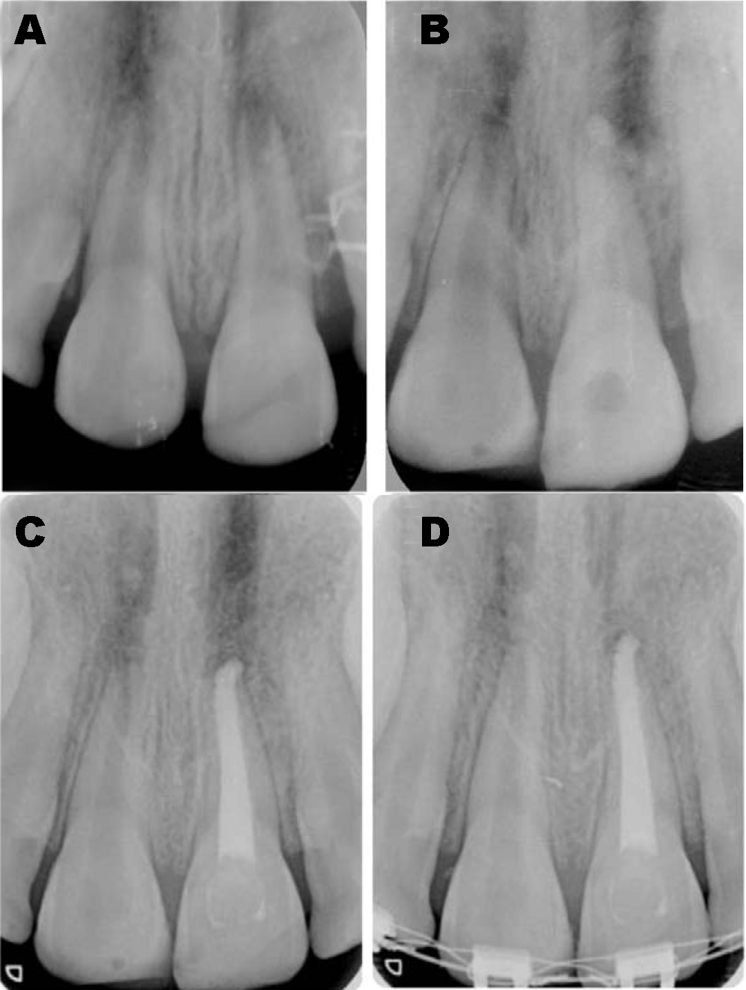
Case 1: A nine-year-old boy who had suffered trauma one year before visiting the pediatric dental clinic of the Faculty of Dentistry of the University of Seville (Spain) due to a change in color and phlegmon formation in the upper left central incisor. The X-ray revealed an open apex in 21, with an apical radiotransparency (Figure 1A). As emergency treatment, the phlegmon was drained surgically intraorally and antibiotic treatment was prescribed (amoxicillin 50 mg/kg daily) during one week. Apexification was started after this week of initial treatment. Under local anesthesia, isolation with a rubber dam was carried out, with chamber aperture of tooth 21. The working length was determined radiographically, and the root canal was carefully cleaned with K-files (Dentsply Maillefer, Ballaigues, Switzerland), without the traditional instrumentation procedure. Between each successive file, we irrigated the root canal with 2.5% sodium hypochlorite, and the canal was posteriorly filled with pure calcium hydroxide powder (Merck, Darmstadt, Germany) dissolved in physiological saline solution, and left in place for one week (Figure 1B). Access to the cavity was sealed with IRM (Dentsply, Germany), leaving a sterile cotton pellet in the pulp chamber. One week later, the calcium hydroxide was removed from the canal aided by carefully instrumentation with K-files (Dentsply Maillefer, Ballaigues, Switzerland) and sodium hypochlorite irrigation, and the root canal was dried with sterile paper tips. We then mixed powdered gray MTA ProRoot (Dentsply Tulsa Dental, Johnson City, TN, USA) with physiological saline solution in 3:1 proportion, and transferred the mixture to the interior of the canal with an amalgam carrier. The material was condensed with blunt paper tips until reaching the apical extremity of the root to form a 3-mm MTA plug. A humid cotton pellet in turn was placed at the entry to the canal to facilitate setting of the MTA, and the cavity was finally sealed with IRM. On the following day conventional endodontic treatment was carried out using gutta-percha (Hygenic, Akron, OH, USA) and AH Plus as sealing cement (Dentsply, Konstanz, Germany), using the lateral condensation technique (Figure 1C); definitive tooth restoration was carried out with composite resin (Enamel Plus HRi, Micerium, Italy). On occasion of the control visit one year later, apical repair was seen to be complete, with rounding of the apex (Figure 1D). |
|
|
|
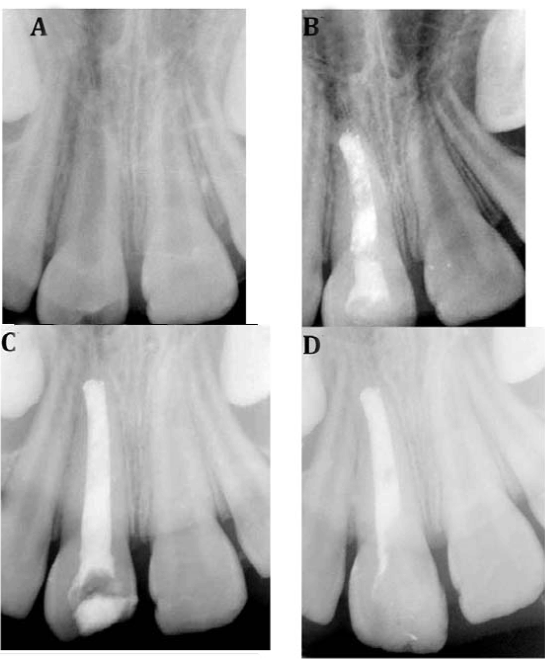
Case 2: An eight-year-old girl visited the pediatric dental clinic due to a change in color, pain and swelling in the upper right central incisor. Direct pulp capping with a temporary restoration had been carried out due to trauma one year before. X-ray revealed an open apex, with an apical radiolucency (Figure 2A). As emergency treatment, antibiotic treatment was prescribed (amoxicillin 50 mg/kg daily) during one week. Following this initial treatment, apexification was carried out in the same way as in the previous patient (Figure 2B-C), though in this case the canal was filled with calcium hydroxide (Merck, Darmstadt, Germany) and Kri-1 (2.025% 661-P-chlorophenol, 4.86% camphor, 1.21% menthol, 80.8% iodoform, 6.5% lanoline and 4.6% glycerin) to afford radiopacity. This material was likewise left in place for one week before applying the gray MTA ProRoot (Dentsply Tulsa Dental, Johnson City, TN, USA). The X-ray after one year showed complete apical repair with rounding of the apex, and no other symptoms (Figure 2D). |
|
|
|
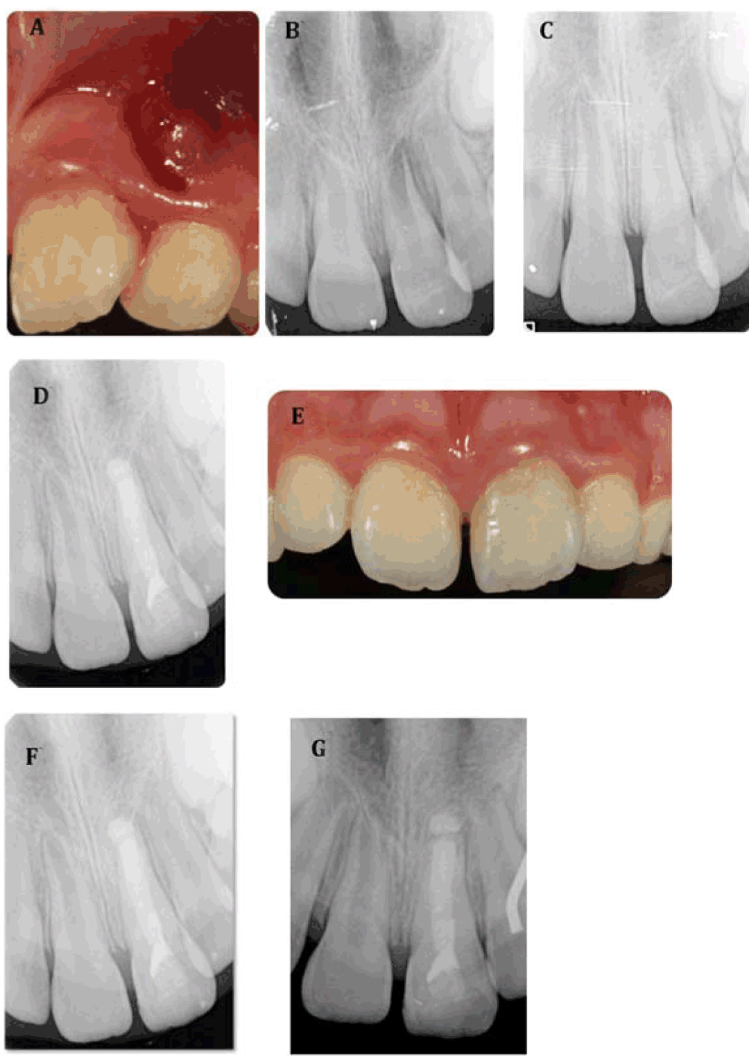
Case 3: An eight-year-old girl visited the pediatric dental clinic due to phlegmon and pain in the upper left central incisor. The parents explained that she had suffered injury three months before, with fracture of the enamel and dentin close to the pulp. The fractured crown fragment had been affixed one week after the accident in another clinic. Clinical examination showed a phlegmon between the upper left central incisor and the upper left lateral incisor (Figure 3A). The X-ray study revealed an open apex and periapical radiotransparency in the upper left central incisor (Figure 3B). As emergency treatment, the phlegmon was drained surgically intraorally and antibiotic treatment was prescribed (amoxicillin 50 mg/kg daily) during one week. One week later, the patient was asymptomatic, and apexification was started following the same protocol as in Case 1 (Figure 3C-D). X-ray, after 6 months, showed apical repair to be complete, with rounding of the apex and the formation of hard tissue above the gray MTA ProRoot (Dentsply Tulsa Dental, Johnson City, TN, USA) (Figure 3E-F). Clinical examination showed the absence of infection. At evaluation after one year, the patient was seen to be asymptomatic and showed good periodontal health (Figure 3G). |
|
|
|
Discussion
|
|
The formation of an apical barrier is necessary in order to fill the root canal system without the risk of overfilling. In this sense, apexification with MTA offers an alternative to conventional treatment with calcium hydroxide [14]. The teeth in our three patients had different degrees of apical aperture and were associated with apical periodontitis. All three showed clinical and radiographic success after MTA treatment. When treating a tooth with necrotic pulp, the main objective is to eliminate bacteria from the root canal system. Since instruments cannot be properly used in teeth with open apexes, disinfection and cleaning of the root canal depends on the chemical action of the sodium hypochlorite used as irrigant and calcium hydroxide used as canal lining material[16]. Some authors use a 1% sodium hypochlorite concentration [17], while others prefer a concentration of 5% [2] [3] . In our cases, sodium hypochlorite was used at a concentration of 2.5%. In all three patients, MTA was used to form an apical plug following one week of calcium hydroxide dressing. This MTA plug facilitates root canal obturation without overfilling. The reason for previously applying calcium hydroxide is to limit bacterial infection in the tooth, since both instrumentation and disinfection are complicated by the unusual architectural characteristics of the canal. Some authors leave the calcium hydroxide in place for two weeks [2] or even four weeks [17] in order to limit bacterial infection. However, in our case and coinciding with other authors [2] [7][9][18], we filled the canals for one week, considering the antimicrobial activity of the material, and with the purpose of avoiding MTA extravasation in the apical zone [17]. A study of the effect of calcium hydroxide upon marginal adaptation of the MTA plug under the scanning electron microscope showed the group pretreated with calcium hydroxide to present smaller gaps than the untreated group, i.e., calcium hydroxide was found to improve marginal adaptation of the MTA [18]. In order to prevent infection of the root canal system following calcium hydroxide placement, it is essential to seal the crown with material affording perfect sealing - i.e., glass ionomer, amalgam or composite. In view of the excellent biological properties of MTA and its capacity to offer good sealing, this material has been recommended for creating an artificial apical barrier in teeth with an open apex - thereby shortening treatment to two or even a single visit [8]. On the other hand, in order to reduce fracture risk, different investigators [8] [19] have proposed apexification in a single visit, placing an MTA apical plug in the last 5 mm of the root canal. Filling of the canal with crown restoration on an immediate basis is therefore possible, and is regarded as a key factor for the long-term preservation of the treated tooth. In our third case, there was slight overfilling. The radiological investigations after six months confirmed apical repair and rounding of the apex. In the case reported by Tahan et al. [17], where excessive MTA extrusion was documented, the subsequent follow-up evaluations showed the patient to remain asymptomatic, and the X-ray study confirmed repair of the periapical lesion. It was therefore concluded that the MTA exerted no deleterious effects. In contrast, Erdem et al. [14], likewise in a case of MTA extrusion, observed no healing of the radiotransparent zone after one year, and a fistula was moreover identified, with increasing tooth mobility that ultimately required extraction. Such overfilling could have been avoided by placing resorbable collagen sponges at apical level, as reported by Bargholz et al. among others [20]. The follow-up X-ray studies in our three patients confirmed bone healing, and the patients were seen to be asymptomatic. The results obtained with MTA are similar to those reported by other authors [2] [19]. |
|
Conclusion
|
|
Mineral trioxide aggregate provided apical stop that enable root canal filling of three teeth in three different clinical cases. |
|
References
|
|

|
[HTML Abstract]
[PDF Full Text]
|
|
Author Contributions:
Asunción Mendoza-Mendoza – Substantial contributions to conception and design, Acquisition of data, Analysis and interpretation of data, Drafting the article, Revising it critically for important intellectual content, Final approval of the version to be published M. Cruz Moreno-Hidalgo – Analysis and interpretation of data, Drafting the article, Revising it critically for important intellectual content, Final approval of the version to be published Carolina Caleza-Jiménez – Analysis and interpretation of data, Drafting the article, Revising it critically for important intellectual content, Final approval of the version to be published Alejandro Iglesias-Linares – Analysis and interpretation of data, Drafting the article, Revising it critically for important intellectual content, Final approval of the version to be published Rosa Yañez-Vico – Analysis and interpretation of data, Drafting the article, Revising it critically for important intellectual content, Final approval of the version to be published Beatriz Solano-Mendoza – Analysis and interpretation of data, Drafting the article, Revising it critically for important intellectual content, Final approval of the version to be published |
|
Guarantor of submission
The corresponding author is the guarantor of submission. |
|
Source of support
None |
|
Conflict of interest
Authors declare no conflict of interest. |
|
Copyright
© 2014 Asunción Mendoza-Mendoza et al. This article is distributed under the terms of Creative Commons Attribution License which permits unrestricted use, distribution and reproduction in any medium provided the original author(s) and original publisher are properly credited. Please see the copyright policy on the journal website for more information. |
|
|