| Table of Contents |  |
|
Editorial
| ||||||
| Molar incisor hypomineralization (MIH): A possible factor in the high prevalence of dental caries in developing nations | ||||||
| Arthur Musakulu Kemoli | ||||||
|
Associate Professor and Chairman, Department of Paediatric Dentistry and Orthodontics, School of Dental Sciences, University of Nairobi, Nairobi, Kenya.
| ||||||
| ||||||
|
[HTML Abstract]
[PDF Full Text]
[Print This Article]
[Similar article in Pumed] [Similar article in Google Scholar] |


| How to cite this article |
| Kemoli AM. Molar incisor hypomineralization (MIH): A possible factor in the high prevalence of dental caries in developing nations. Edorium J Dent 2015;2:51–55. |
|
Dental caries
| ||||||
|
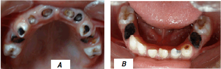
Dental caries is as old as mankind, and even during the Roman occupation of Europe in the fourth and fifth century, there was a general increase in dental caries in European population to an all time level of 71.6%. The increase was thought to be associated with increased consumption of cooked foods that included bread, porridge of cereals, vegetables, olives, some fruits and wine, meats [1][2]. The refinement and the cooking of the foods gave rise to increased retentive and sticky effects with low oral clearance times, [3] [4], potential precursor to the development of dental caries. As late as 1970s, the prevalence of dental caries in Europe and other western nations had still continued to rise. The increase was attributed to further dramatic increase in the intake of sugar and refined carbohydrates. This trend was conspicuously absent in the developing nations, [5]. In the past few decades, the global trend of dental caries has shown a reversal of the previous trend, where the developing nations are seemingly groping with increasing prevalence of dental caries while the western developed nations are now enjoying declined or plateaued trends in respect to the prevalence of dental caries, particularly in the child-population. The situation in the developed nations has been helped by the aggressive dental public health measures, improvement in dental treatment, widespread use of fluoride toothpaste and fluoridated drinking water, and the changing social factors with improvement in the general health indicators in these nations [6]. On the other hand, the increasing affluence and urbanization in the developing nations, coupled with deficient oral health education, malnutrition, absence of health services and poor quality of life have enhanced the prevalence of caries [7] to such high levels in children (Figure 1). The development of Venn diagram in the 1960s, depicted the aetiological factors of dental caries as composed of the tooth, diet and cariogenic bacteria, helped the clinicians to understand more about the disease. However, in the fullness of time, many other modifiers of the process have become to be recognized [8]. These modifiers have included dental plaque fluids, saliva pH, presence of calcium/phosphate ions, use of fluorides, immune system, time, socioeconomic status, level of education, lifestyle and behavioral factors. The interplay of these factors/modifiers provided further insight to the dynamism of the carious process that has now been recognized as involving a demineralization-remineralization process of the tooth hard tissues. The maintenance of the stability of this equilibrium has become to be the best approach to the long-term management of dental caries. | ||||||
| ||||||
|
Molar incisor hypomineralization
| ||||||
|
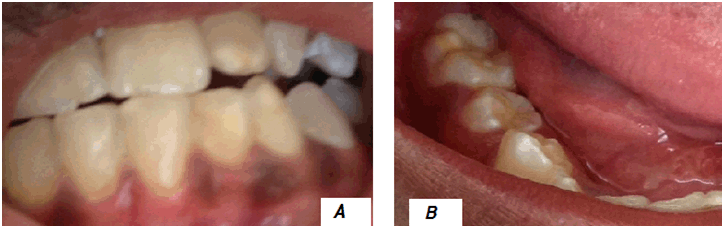
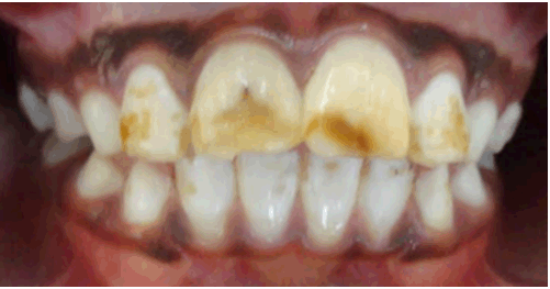
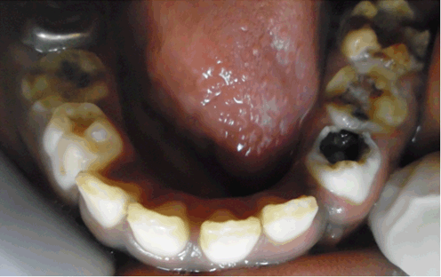
Molar incisor hypomineralization (MIH) is another dental problem that has recently been taking a centre stage in dentistry, especially in the child-population. While its aetiology is not very clear-cut [9], it is known to be a disturbance to the dental enamel during the stage of odontogenesis, during which time, the resorption potential of the ameloblasts appear to get affected through the inhibition of proteolytic enzyme, resulting in protein retention, interference with enamel crystal growth and the enamel maturation [10] [11][12][13]. MIH afflicts the permanent first molars with or without the permanent incisors and occasionally the second primary molar, resulting in areas of enamel hypomineralization of these teeth. The hypomineralised areas are seen as opacities within the enamel, and are limited to the incisal or cuspal one-third of the dental crown and will rarely involve the cervical one-third of the tooth [13] (Figure 2). The putative factors associated with molar incisor hypomineralization have, therefore, included systemic conditions and environmental insults during the early development of teeth especially in the period after birth and three years of age [14]. These conditions include poor general health, exanthematous infections, nutritional diseases, nutritional deficiencies, systemic diseases, high fevers, infections, toxins, radiation, brain injury and neurologic defects, cystic fibrosis, syndromes of epilepsy and dementia (Kohlschutter-Tonz syndrome), nephrotic syndrome, atopia, lead poisoning, repaired cleft lip and palate, radiation treatment, rubella embryopathy, epidermolysis bullosa, ophthalmic conditions, celiac disease, renal conditions, pre-term births, gastrointestinal disorders and possibly genetics [15][16][17]. Although fluoride is a risk factor for MIH, being usually associated with generalized diffuse defects of enamel (Figure 3), the evidence to its involvement with the aetiology of MIH is weak [18][19]. Just as in some cases of dental caries, a clinical examination can be used to arrive at a diagnosis of MIH. The affected tooth will show demarcated opacities with a discoloration that ranges from white to yellow-brown [12]. These demarcated opacities are susceptible to post eruption breakdown (PEB), and even after restoration, the affected teeth will show atypical restorations due to continued breakdown of the tooth structure [20], that may generally results in early extractions due to severe caries-involved pulpal tissue. The MIH affected teeth, the first permanent molars and/or permanent incisors, are often hypersensitive to temperature changes and difficult in achieving local anesthesia, thus exposing the child to a neglected oral health with subsequent early loss of the dentition (Figure 4). | ||||||
| ||||||
| ||||||
| ||||||
|
Dental caries-MIH interplay
| ||||||
|
The prevalence of dental caries in the developing nations has been on the rise in the last few decades, and there are indications that the cases of MIH could also be rising, going by the few studies already done in the developing nations [21][22][23][24][25]. It is possible that the presence of MIH could be contributing to the rise in the prevalence of dental caries in children in the developing nations. Considering that an erupting permanent first molar afflicted by MIH (associated with poor nutrition) is highly susceptible to dental caries and erosion, and given that the majority of the children in developing nations come from poor families with poor nutritional status, the possibility that this tooth is at risk to dental caries is very high. This situation is even made worse with the increasing urbanization, changes in the living conditions and lifestyles, increasing availability and access to processed foods and sweet drinks to the children [26], promotive factors to dental caries. Dietary practices and nutrition are undergoing changes in most developing countries and the consumption of sugars is increasing rapidly among the children in these nations. [27][28] Hence, early childhood caries is becoming more prevalent, and most likely MIH as well [29]. The urbanization, coupled with globalization of the children living in the developing nations is supposed to be the best thing to happen to these children, as it provides them or their parents with free access to healthcare information and the availability of diverse foods to most children. However, all these changes are coming at a price in the form of the sharp rises in the prevalence of dental caries [30], and probably MIH. Coupled with all these are the socio-economic determinants in these nations where children have reduced oral self-care practices, attitudes and values towards oral health and even poor diet and nutrition [31]. All these determinants and social exclusions have real bearing to the etiology of dental caries and MIH [32]. A quick look at the global prevalence of MIH in children reveals a much varied situation in its prevalence, that ranges from 2.8% to 40.2% [18][19] [33]. The highest figures have in fact been recorded in Brazil and Denmark [34]. Some of the developing nations that have reported prevalence of MIH have been Libya (9%), Kenya (13.7%) and Brazil (40.2%) [21] [25]. These few studies appear to show that MIH could be a significant problem in these nations, particularly when coupled with poor oral hygiene and nutritional status, a panacea to dental caries. Given the fact that MIH is associated with structural weakness of the tooth and hypersensitivity, there exists a very high chance of the children afflicted with MIH will be unable to maintain good hygiene and therefore experience dental caries. The fact that histological examination of hypomineralized teeth has previously shown that oral bacteria can get embedded deep into the dentine of the affected teeth [35], there is an obvious chance of increased risk to dental caries to the affected teeth. From a foregoing statements, it can be inferred that, although the cases of dental caries are rising in the developing nations due to associated poverty and restricted diet (rich in carbohydrates and poor in animal protein) and facilitated frequent intake of cariogenic foods due to some periods of better economic conditions. Besides, defective dental structure development due to malnutrition and hypocalcification can increase the vulnerability of the dentition to caries attack [36]. The dental practitioner today should, therefore, take cognizant of this changing pattern in the development of dentition in children and environmental conditions, and be ready to provide intensive dental caries and MIH preventive measures, particularly for those children living in developing nations. Thorough oral hygiene should be a priority, and should include the promotion of dental remineralization through the use of fluoridated toothpastes, fluoride varnishes and other supplements. In the case of MIH, the remineralization therapy should commence as soon as the defective surface is accessible. The use of casein phosphopeptide-amorphous calcium phosphate could prove good for these teeth. The potentially more costly and more time consuming approach, of the provision of sealants could augment the other measures, and more so in the prevention of occlusal caries on permanent first molars. The application of the cheaper ART glass-ionomer cement sealants using the finger-press technique can also come in handy, especially when the teeth are not fully erupted [19]. However, it should also be noted that in MIH afflicted teeth, when PEB occurs, the porous subsurface enamel or dentin becomes exposed, resulting in tooth sensitive temperature changes and tooth-brushing. Poor oral hygiene favors plaque retention and promotes rapid caries development [37] [38]. In this case, urgent attention to restore the tooth either with use of a dental restorative material or a dental crown has to be considered. At the very worst, extraction of the permanent first molars can be an alternative where the erupting permanent secondary molars are in good position to take the place of the extracted molars, or an extraction followed by appropriate prosthesis can be an option. It is instructive, therefore, for the dental practitioner to know that the rising childhood dental caries in the developing nations and the apparent increasing cases of MIH may have more factors coming into play than the known traditional ones [39] [40]. Children living in the developing nations face multitude of problems, some of them are etiological factors for dental caries and MIH. Other factors; these children are exposed to make it harder for them to maintain sustainable oral health preventive measures. The maldistribution of oral health personnel in developing nations means children living in some parts of these nations have less professional support, fewer choices and opportunities for specialized oral health care. Discrepancies or non-existence of oral health insurance coverage to these children can only worsen the situation [41] [42]. It would help if oral health were to be fully integrated into primary health care (PHC) services, and that the PHC workers, who are mostly non-dental health care providers and who are more accessible in rural areas, provide basic oral health information and screening. [43] The cadre of health workers can help in early diagnosis of dental caries and MIH. They can also help promote the efficient ways of disrupting the oral microbial homeostasis in the 'biofilm' and also the disruption of the mineral homeostasis between the tooth and the 'oral fluid'. This will in effect help protect the normal or MIH-afflicted teeth from dental caries. Keywords: Dental caries, Dental plaque fluids, Immune system, Molar incisor hypomineralization (MIH) | ||||||
|
References
| ||||||
| ||||||

|
[HTML Abstract]
[PDF Full Text]
|
|
Author Contributions:
Arthur Musakulu Kemoli – Substantial contributions to conception and design, Acquisition of data, Analysis and interpretation of data, Drafting the article, Revising it critically for important intellectual content, Final approval of the version to be published |
|
Guarantor of submission
The corresponding author is the guarantor of submission. |
|
Source of support
None |
|
Conflict of interest
Authors declare no conflict of interest. |
|
Copyright
© 2015 Arthur Musakulu Kemoli. This article is distributed under the terms of Creative Commons Attribution License which permits unrestricted use, distribution and reproduction in any medium provided the original author(s) and original publisher are properly credited. Please see the copyright policy on the journal website for more information. |
|
|