| Table of Contents |  |
|
Original Article
| ||||||
| Root thickness evaluation of mandibular incisors | ||||||
| Nahid Mohammadzadeh Akhlaghi1, Bahareh Dadresanfar2, Pooneh Mohebbi2, Mehdi Vatanpour2, Shabnam Sohanian3 | ||||||
|
1Associate Professor, Endodontic Department, member of cranio-maxillo-facial Research center, Dental Branch, Islamic Azad University, Tehran, Iran.
2Assisstant Professor, Endodontic Department, Dental Branch, Islamic Azad University, Tehran, Iran. 3Post-graduate Student, Oral and Maxillofacial Pathology Department, Babol, Mazandaran, Iran. | ||||||
| ||||||
|
[HTML Abstract]
[PDF Full Text]
[Print This Article]
[Similar article in Pumed] [Similar article in Google Scholar] |


| How to cite this article |
| Akhlaghi NM, Dadresanfar B, Mohebbi P, Vatanpour M, Sohanian S. Root thickness evaluation of mandibular incisors. Edorium J Dent 2015;2:56–60. |
|
Abstract
|
|
Aims:
Mandibular incisors are the smallest teeth in the mouth. They have thin roots with concavities and are very important for aesthetic and also for speaking. Due to their narrow internal anatomy, preparing the root canal and post space might be very difficult and endangers them for root wall perforation or fracture. The aim of this study was to measure the root thickness of mandibular incisors.
Methods: Root wall thicknesses of eighty mandibular incisors were measured in four sections including the CEJ, 3 mm apical to the CEJ, 4 mm and one mm coronal to the apex and in buccal, lingual and proximal surfaces of concavity areas, using a stereomicroscope. Data were statistically analyzed by repeated measured ANOVA and paired t-test. Results: Buccal surface of the buccal canals and lingual surface of the lingual canals in double-canalled incisors had the greatest root thicknesses compare to the proximal surfaces. Root thickness of single-canalled root was more than of double-canalled ones. Proximal walls had the least root thicknesses in all the sections especially in section 4, at 1 mm from the apex, which was less than 1 mm. Conclusion: In order to avoid technical mishaps during root canal procedures, attention must be paid to the thin concavity proximal walls using anticurvature flaring and also in selection of proper size of master apical file. | |
|
Keywords:
Mandibular incisors, Root, Teeth, Thickness
| |
|
Introduction
| ||||||
|
The amount of residual dentin is crucial to the survival of a root filled tooth [1]. To avoid iatrogenic mishaps during procedures such as canal instrumentation and preparing the dowel space, one should have enough knowledge about the radicular dentin thickness. Considerable dentin loss, especially where grooves are present or between two fused roots, can initiate periodontal involvement that are difficult to manage [2]. In addition to periodontal problems, it has been proposed that dentin thickness in association with canal curvature and external root morphology are factors potentially influencing fracture susceptibility [3]. The thinner the dentin, the greater is the likelihood of tooth fracture [4]. In a study performed by Katz et al, it was reported that in roots with less mesiodistal dimension than the buccolingual, such as maxillary and mandibular premolars, mesial roots of mandibular molars and mandibular incisors, the risk of fracture due to root canal and post space preparation is greater [5]. As mandibular incisors have the smallest size among the teeth [6], the importance of having enough knowledge regarding their thickness is high lightened. Little information is available about the thickness of different canal walls in these teeth before preparation procedures [1]; so the aim of this study was to measure the radicular root thickness in mandibular incisors. | ||||||
|
Materials and Methods
| ||||||
|
One hundred and forty human mandibular incisors, extracted due to periodontal problems, were collected from Iranian subjects aged between 35 and 55 years, after the study had been approved by the ethics committee of Dental Branch, Islamic Azad university of Tehran (Ethics committee approval 10001). After immersing the samples in 5.25% NaOCl for 24 hours, they were cleaned of calculus with an ultrasonic instrument. The teeth with root caries, cervical or apical resorption, open apices or fractures were excluded. The root length was measured from the buccal CEJ to the apex by a ruler. Then the access cavity was prepared and a #10 K-file (Dentsply, Maillefer, Tulsa, OK) was inserted until it was visible at the apical foramen. A parallel radiographic technique with an E-speed film (Kodak, Stuttgart, Germany) was used for each tooth after stabilizing it with red molding wax in a buccolingual direction. The exposure time and source-to-film distance was the same for all samples (0.6 s; 70 kVp; 20 cm). Teeth with prior root canal therapy and internal resorption were excluded. All the radiographs were scanned using an hp scanner (Canoscan 3200 F; Canon, Tokyo, Japan). Images were processed using Autocad 2002 (AutoDesk, San Rfel, CA, USA) and canal curvature was determined based on Schneider technique [7]. The root surfaces of all the samples were stained with methylene blue and then invested in clear acrylic blocks; the buccal surfaces were marked by a vertical groove. The teeth were sectioned in four horizontal parallel planes perpendicular to the long axis of each tooth:
All the measurements were carried out with the help of an accurate mechanical caliper using a diamond-coated disk (D & Z, Germany) with a thickness of 0.1 mm and a diameter of 22 mm. Each tooth was coded and the canal orifices of each section were stained with fuchsin after fixing them on a glass slide. A stereomicroscope (Olympus, SZX-ILLB200, Japan) was used for the root width measurements under ×12.5 magnification. Images were captured by a digital camera and saved in a computer. A total of 80 mandibular incisors including 20 double-canalled and 20 single-canalled incisors from subjects aged 35–45 and 20 double- and 20 single-canalled from subjects aged 46–55 remained in the study. For each section, the following root surfaces were evaluated for thickness: buccal (distance from the buccal wall limit of the canal to the outer surface of the root); lingual (distance from the lingual wall limit of the canal to the outer surface of the root); proximal (from the mesial/distal wall limit of the canal to the outer surface of the root). In addition to all the above-mentioned measurements in double-canalled roots, concavities of the buccal and lingual canals in the proximal areas were measured. All the measurements were made to 0.01 mm accuracy using Adobe Photoshop 7.0 software. Data were recorded and statistically analyzed by repeated measures ANOVA and paired t-test. | ||||||
|
Results | ||||||
|
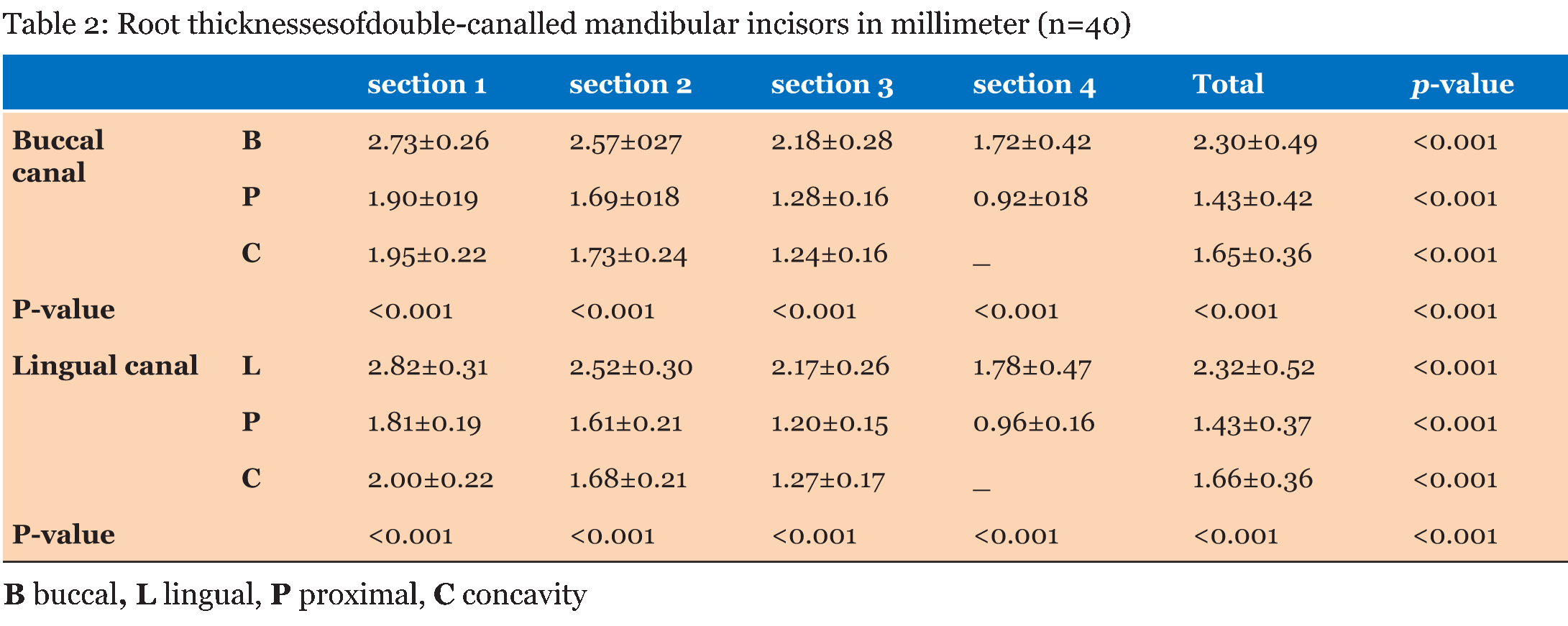
The average of root lengths was 13.08 mm and 13.85 mm for double-canalled and single-canalled roots, respectively. The average of canal curvature was 8° and 16.88° in double-canalled and single-canalled roots, respectively. Correlation coefficient did not show any significant relationship between canal curvature and root length and radicular thickness. Root thickness mean values and standard deviations for each section of the samples were listed in Table 1 and Table 2. Lingual and buccal surfaces had the maximum root thicknesses and the proximal surfaces had the minimum root thicknesses (p<0.001). At 4 mm and 1 mm coronal from the apex the proximal surface (mesial/distal) exhibited significant differences from other surfaces. Repeated measures ANOVA was used to compare root thicknesses in each of the four sections; there were significant differences among them at each surface (p<0.001). Two-by-two comparisons of buccal, lingual and proximal surfaces at each section showed significant differences, with root thickness values decreasing toward the apex (p<0.001). Single-canalled and double-canalled comparisons Root thickness comparison in different age groups
| ||||||
| ||||||
|
| ||||||
| ||||||
|
Discussion
| ||||||
|
In this study, histological cross sections of extracted teeth were investigated. In order to prevent root fracture and record the position of the walls in each section, the teeth were invested in clear acrylic resin [8]. A stereomicroscope and a digital camera were used to take photomicrographs and save them in a computer because of their availability and convenience. In other studies, stereomicroscopes, hp scanners, electron and light microscopes, digital cameras and computed tomography techniques have been used [9][10][11][12][13]. As root thickness is affected by age, an age range of 35–55 was selected and studied in two groups: 35–45 and 46–55 years of age. Root thicknesses were determined in four sections. In previous studies, one [10] , two [14], three [5] , four [13] [15] or even more [11] sections have been evaluated based on the aim of the study. Some studies have only reported the average value in each section and not the maximum and minimum wall thickness separately [1] [11][14][15]. In other studies, residual root thickness after dowel space preparation has been reported, without reporting the initial root thickness [10][15]. The results of the present study showed that the lingual and buccal surfaces had the maximum root thickness while the proximal surfaces had the minimum thickness (p<0.001). Therefore, in cleaning and shaping of these canals the dental practitioner should avoid removing too much dentin from the proximal walls. Minimum root thickness of proximal surfaces can be attributed to the longitudinal axis of teeth in the dental arch, and also the functional and occlusal forces that can affect the thickness of buccal and lingual surfaces. In other studies [1] [5], similar to the present study, the thickest root wall was the lingual, but the reported thicknesses were different, which might be attributed to the evaluation of mandibular incisors collectively and not separating them into single- and double-canalled roots or evaluation of different societies with different ethnic backgrounds. At 4 mm from the apex, proximal root thickness was almost 1.2 mm; therefore, there is a risk for thinning and perforating the root while preparing the dowel space, which is consistent with the results of other studies [1] [6][9][16] reporting that the root surfaces at the apical third were the thinnest in all the anterior teeth. At one millimeter from the apex, proximal root thickness was less than 1 mm, which is less than the minimum thickness for preserving the root [17]. Therefore, it is important to select a suitable master apical file. This finding is consistent with those of other studies [1] [14]. Murray et al. [13] showed that the thicker root surfaces of radicular sections were at the apical quarter in the oldest age group (51–59) in comparison to 31–50 age group. In the present study root thicknesses in all the sections were greater in the 46–55 age group but the differences from the 35–45 age group were not significant. | ||||||
|
Conclusion
| ||||||
|
Based on the results of the present study, root thickness of double-canalled and single-canalled incisors at 4 mm from the apex was less than 1.5 mm and 1.2 mm respectively. Therefore, it would be logical to finish dowel space somewhere coronal to the 4 mm from the apex. In addition, considering thin root thickness of proximal surfaces, dentin removal should be directed to thick walls of the buccal and lingual aspects. Care should be taken during instrumentation of mandibular incisors, especially in the mesiodistal aspect to avoid perforation, which is most likely to occur at the apical third. | ||||||
|
References
| ||||||
| ||||||


|
[HTML Abstract]
[PDF Full Text]
|
|
Author Contributions:
Nahid Mohammadzadeh Akhlaghi – Substantial contributions to conception and design, Acquisition of data, Drafting the article, Revising it critically for important intellectual content, Final approval of the version to be published Bahareh Dadresanfar – Substantial contributions to conception and design, Acquisition of data, Drafting the article, Revising it critically for important intellectual content, Final approval of the version to be published Pooneh Mohebbi – Substantial contributions to conception and design, Acquisition of data, Drafting the article, Revising it critically for important intellectual content, Final approval of the version to be published Mehdi Vatanpour – Substantial contributions to conception and design, Acquisition of data, Drafting the article, revising it critically for important intellectual content, Final approval of the version to be published Shabnam Sohanian – Substantial contributions to conception and design, Acquisition of data, Drafting the article, Revising it critically for important intellectual content, Final approval of the version to be published |
|
Guarantor of submission
The corresponding author is the guarantor of submission. |
|
Source of support
None |
|
Conflict of interest
Authors declare no conflict of interest. |
|
Copyright
© 2015 Nahid Mohammadzadeh Akhlaghi et al. This article is distributed under the terms of Creative Commons Attribution License which permits unrestricted use, distribution and reproduction in any medium provided the original author(s) and original publisher are properly credited. Please see the copyright policy on the journal website for more information. |
|
|