| Table of Contents | |
|
Case Report
| ||||||
| Focal cemento-osseous dysplasia of mandibular jaw: A case report and literature review | ||||||
| Shahabe Saquib Abullais1, Priyanka2, Nitin Kudyar3, G. Kapil4, Prashanth Shetty5 | ||||||
|
1Assistant Professor, Department of Periodontics, King Khalid University, Abha, Saudi Arabia.
2Assistant Professor, Department of Periodontics, Yogita Dental College and Hospital, Khed, Ratnagiri, Maharashtra. 3Associate Professor, Department of Periodontics, Himachal Dental College and Hospital, Sundarnagar, Himachal. 4Assistant Professor, Department of Endodontics, Yogita Dental College and Hospital, Khed, Ratnagiri, Maharashtra. 5Associate Professor, Department of Periodontics, Yogita Dental College and Hospital, Khed, Ratnagiri, Maharashtra.. | ||||||
| ||||||
|
[HTML Abstract]
[PDF Full Text]
[Print This Article]
[Similar article in Pumed] [Similar article in Google Scholar] |
| How to cite this article |
| Abullais SS, Priyanka, Kudyar N, Kapil G, Shetty P. Focal cemento-osseous dysplasia of mandibular jaw: A case report and literature review. Edorium J Dent 2015;2:61–64. |
|
Abstract
|
|
Introduction:
Focal cemento-osseous dysplasia is a rare disease entity characterized by replacement of healthy bone and tissue with cemento-osseous tissue. Etiology and pathogenesis for the lesion is unknown and these lesions are considered as reactive lesion in the periapical areas of the teeth.
Case Report: A 52-year-old female reported to Yogita Dental College and Hospital with the chief complaint of throbbing intermittent pain and swelling in the lower left posterior region of the jaw since six months. Conclusion: The lesions are benign in nature and rarely cause complication. Most of the time lesion is detected accidently on the routine radiographs. Treatment involves long-term follow-up of the case unless until lesion becomes symptomatic. | |
|
Keywords:
Alveolar bone, Benign lesion, Cementum, Focal cemento-osseous dysplasia
| |
|
Introduction
| ||||||
|
Cemento-osseous dysplasias are group of disorders known to originate from periodontal ligament tissues. They are rare entities which are characterized by replacement of healthy bone and tissue with cemento-osseous tissue [1]. These lesions are benign and related to tooth bearing areas of the jaw bone. They are usually classified, into three groups [2]:
The term cemento-osseous dysplasia was adopted for the WHO classification in 1992. It is difficult to discriminate cementum from bone tissue in lesions which produce cementum, bone and connective tissue, which is why lesion has been named cemento-osseous dysplasia. Focal cemento-osseous dysplasia (FCOD) is accepted as most frequently seen fibro-osseous pathology [3]. The FCOD is predominantly seen in African-American females and less in African-American males, with a peak incidence in the fourth and fifth decades of life [4] [5]. The FCOD can cause localized expansion of the alveolar bone and can be secondarily infected with localized abscess formation [6]. The etiology and pathogenesis for FCOD is unknown, and this lesion is considered as a reactive lesion in the periapical areas of the teeth. Often it affects one or two mandibular anterior tooth. Involvement of mandibular posterior teeth is also reported [3]. When it involves single tooth, in the initial stages, clinician may get confused with periapical granuloma or cyst. The FCOD is often a well defined radiolucency with a sclerotic border or a mixed radiolucent and radiopaque lesion. We can arrive at a provisional diagnosis of cemento-osseous dysplasia in cases when there is absence of pain or swelling and the presence of a vital pulp in the affected portion of the jaw, coupled with the typical clinical and radiographic findings for this condition. When a diagnosis of FCOD is established, periodic recall is recommended. The lesion may persist for indefinite period of time without causing any symptoms. No surgical intervention is required for the asymptomatic patient. For symptomatic cases or when the lesion is secondarily infected, surgical intervention is recommended [7]. | ||||||
|
Case Report
| ||||||
|
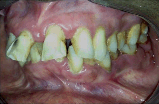
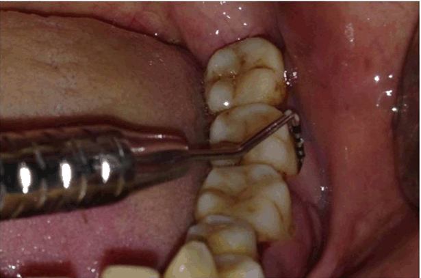
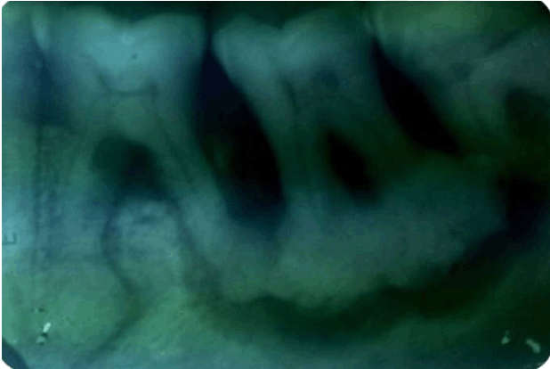
A 52-year-old female reported to Yogita Dental College and Hospital with the chief complaint of throbbing intermittent pain and swelling in the lower left posterior region of the jaw since six months. Pain got aggravated after food lodgment and relieved after removal of stimulus by tooth pick or by taking NSAID. The patient was questioned for relevant systemic diseases, history of smoking and past dental history. Intraoral examination revealed hard non-tender swelling in the lower left posterior region of the jaw with obliteration of the buccal vestibule (Figure 1). Periodontal examination revealed deep periodontal pocket with 36 and 37 (Figure 2). Surprisingly, the affected teeth in spite of severe attachment loss did not show significant mobility. Vitality test of the affected teeth revealed positive response. The IOPA of lower left posterior region revealed a well defined mixed radiolucent radiopaque lesion centered over the periapical region of 37 and also involving the apices of both the roots of 36 (Figure 3). The borders of the lesion were sclerotic and the lesion had a roughly oval shape. The internal structure appeared as an amorphous, well defined radiopaque mass surrounded by a well defined radiolucent periphery. A second similar but smaller lesion was seen attached to the mesial root of 36. Both the lesions were separated from each other by bony trabeculae which appeared to be normal in density. On the basis of clinical findings and radiographic examination, a provisional diagnosis of FOCD was given. Histopathological evaluation was done for confirmation of the clinical and radiologic findings. A small section of diseased tissue was obtained through incisional biopsy for histopathological evaluation. Involved teeth were not extracted as per the request of the patient. The hematoxylin and eosin stained and studied section of the lesional tissue showed presence of dense lamellar bone with presence of typical resting lines. The bony trabeculae did not show presence of any osteoblastic rimming (Figure 4). | ||||||
| ||||||
| ||||||
| ||||||
| ||||||
|
Discussion
| ||||||
|
Cemento-osseous lesions are a result of disturbance in the bone metabolism which results in replacement of normal bone with the connective tissue matrix that then gradually develops in to cemento-osseous lesion in the later stage of the disease. Waldron reported the first case of FCOD as 'localized fibro-osseous cemental lesion [3]. Later, Summerlin and Tomich renamed it as focal cemento-osseous dysplasia [8]. The FCOD clinically and histopathologically mimics ossifying fibroma and imposes a difficulty in diagnosis. Ossifying fibromas are well-demarcated and show radiolucent feature with small radio-opaque calcifications. I it is easy to excise because of well demarcated radiolucent border. However, FCOD is usually radio-opaque and cannot be readily excised from healthy bone [6]. The FCOD is usually seen in asymptomatic patients in the periapical area of teeth with vital pulp. A few cases have been reported with localized jaw expansion and mild discomfort. The age of occurrence is in fourth or fifth decade of life with higher predilection in females [9]. The patient in the present case was also a female in her fifth decade of life. The patient was complaining of throbbing pain, mild discomfort and expansion of jaw in the 36 region. Most of the cases of FCOD are reported accidently while shooting routine radiographs. Seventy percent of the FCOD cases are associated with root apex of the teeth. The remaining 21% is found in the previous extraction site. In presented case radiograph was taken to find out the cause of food impaction and discomfort to the patient and accidentally the lesion was found in the periapical area. The FCOD has three developmental stages with specific radiographic features;
| ||||||
|
Conclusion
| ||||||
|
There are wide varieties of periapical pathosis present and they resemble each other in many aspects. It is important to know specific clinical, radiographic and histological feature of particular lesion so as to avoid confusion in final diagnosis. The FCOD is rarely diagnosed according to the literature. Thus, due to the importance of FCOD, the lesion must be included during the evaluation of intra-osseous lesions. If the case is asymptomatic treatment is not required, whereas in symptomatic cases surgical excision is advised. | ||||||
|
References
| ||||||
| ||||||
|
[HTML Abstract]
[PDF Full Text]
|
|
Author Contributions
Shahabe Saquib Abullais – Group1 - Substantial contributions to conception and design, Analysis and interpretation of data, Drafting the article, Revising it critically for important intellectual content, Final approval of the version to be published N. Priyanka – Acquisition of data, Analysis and interpretation of data, Revising it critically for important intellectual content, Final approval of the version to be published Nitin Kudyar – Acquisition of data, Analysis and interpretation of data, Revising it critically for important intellectual content, Final approval of the version to be published G. Kapil – Analysis and interpretation of data, Revising it critically for important intellectual content, Final approval of the version to be published Prashanth Shetty – Analysis and interpretation of data, Revising it critically for important intellectual content, Final approval of the version to be published |
|
Guarantor of submission
The corresponding author is the guarantor of submission. |
|
Source of support
None |
|
Conflict of interest
Authors declare no conflict of interest. |
|
Copyright
© 2015 Shahabe Saquib Abullais et al. This article is distributed under the terms of Creative Commons Attribution License which permits unrestricted use, distribution and reproduction in any medium provided the original author(s) and original publisher are properly credited. Please see the copyright policy on the journal website for more information. |
|
|