 |
|
Review Article
| ||||||
| Dental alterations in sickle cell anemia | ||||||
| Antonio Fabrizio Nifosí1, Pablo Castelló2, Lorenzo Nifosí2, Gianfilippo Nifosí3 | ||||||
|
1Doctor of Dental Surgery, Dental Clinic “iDental”, Valencia, Spain
2Student of Dental Surgery, European University of Valencia, Valencia, Spain 3Medical Doctor, Hemato-Oncology Clinic, Brugmann University Hospital Center, Brussels, Belgium | ||||||
| ||||||
|
[HTML Abstract]
[PDF Full Text]
[Print This Article]
[Similar article in Pumed] [Similar article in Google Scholar] |


| How to cite this article |
| Nifosí AF, Castelló P, Nifosí L, Nifosí G. Dental alterations in sickle cell anemia. Edorium J Dent 2017;4:19–23. |
|
ABSTRACT
|
|
The epidemiology of the dental disease in sickle cell anemia is not adequately described. The aim of this work is either the evaluation of literature on the topic, or to describe if the disease itself predisposes to a dental pathology, and eventually also if the latter can influence its course. We selected many cases and reviews in order to identify the dental diseases related to sickle cell anemia. Caries and periodontitis are not directly caused by disease. However, an inflammatory state caused by a dental infection can trigger or precipitate a sickle crisis. Malocclusion angle class II is particularly frequent, as a result of bone facial changes and/or related to muscle imbalance. Temporomandibular joint disorder is possible. Asymptomatic pulp necrosis is due to the sickling that causes vasocclusion within microcirculation of the dental pulp. Large population-based studies are needed in order to clarify the dental involvement in this disease. A strict collaboration between the dentist and the hematologist is essential. | |
|
Keywords:
Hemoglobinopathy, Sickle crisis, Dental tissues, Oral health
| |
|
INTRODUCTION
| ||||||
|
Sickle cell anemia (SCA) is an hereditary hemoglobinopathy, characterized by the production of an abnormal hemoglobin (Hb S), that precipitates in conditions of reduced oxygen tension, forming complexes that deform the red blood cells which leads to a characteristic sickle shape, responsible of vascular occlusion and ischemic injury of many organs and tissues. The most common genotypes observed are HbS/ß0-thalassemia (ßsß0) and HbS/ß+-thalassemia (ßsß+), while the homozygous sickle cell anemia (ßsßs) is more frequent, and the double heterozygous Hb S/HbC (ßsßc) is rare. It represents a major public health problem following the continuous migration of populations. Chronic hemolytic anemia, sickle crisis and infections are the most frequent manifestations. The head and neck is involved at various levels, including the teeth. Some studies have examined the relationship between oral health and SCA. Dentists play an important role in preventing complications and improving the quality of life of these patients. | ||||||
|
METHODS
| ||||||
|
Systematic critical review of the reports and other reviews are published on the topic in PUBMED, GOOGLE SCHOLAR and LILACS electronic databases, using the following keywords: “dental alterations in sickle cell anemia” and “oral health in sickle cell anemia”. | ||||||
|
RESULTS | ||||||
|
Dental abnormalities | ||||||
|
Several alterations of the dental tissue structures in SCA are described in literature, but there are few studies that illustrate the prevalence of it [1]. A delay in the dental eruption is commonly reported as a result of ischemic bone infarcts, with a 1.7 higher risk compared to the general population [2]. Hypodontia has been described in one patient with Hb SC disease [3]. Abnormal tooth formation includes hypomaturation and hypomineralization of enamel and dentin as responsible of an intrinsic opacity of teeth in 67.5% of the patients, both heterozygous and homozygous patients [4]. Hypercementosis is a consequence of the non-neoplastic cementum excessive deposition during odontogenesis [5]. Infractions are micro-cracks within the enamel, reported anecdotally. Erosions of enamel, more rarely of dentin, are the result of the effects of the acidity on dental tissues. They occur most often in children who drink fruit juices or in adult with certain disorders such as a gastroesophageal reflux [6]. Tooth discoloration maybe the consequence of the incorporation. In the enamel and dentin of endogenous (bilirubin, hemoglobin) and exogenous pigments (medications) [7].
| ||||||
| Caries | ||||||
|
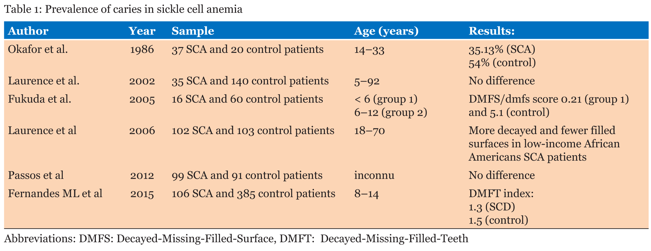
Although there is no direct evidence that sickle cell disease causes the appearance of caries, the SCA patients have increased susceptibility to caries, with a higher prevalence of tooth decay and lower of filled teeth [6][7]. The frequent use of soft drinks containing sugar for hydratation, inadequate oral hygiene and lowest socioeconomic status are predisposing factors [7]. In literature, few studies report the prevalence of caries in SCA patients with contradictory results (Table 1). Other than the disease itself, older age, female gender and daily smoking are the major risk factors of caries also in SCA patients [8]. The severity of the lesions is related to the age, the number of hospitalizations and the need for transfusions or drugs. Regular dental consultation due to frequent hospitalization for sickle crisis makes that in the literature is reported a low caries incidence that subjects without SCA. In sickle children is described a lower prevalence of caries [9], as a result of an acquisition delay of Streptococcus mutans for the prolonged prophylactic penicillin, both also for the oral colonization by cariogenic strains of C. albicans [10]. Socioeconomic factors have been shown to influence caries risk [11]. An increased risk of decayed teeth is not associated with the level of education of the parents but with the low family income [12]. Another negative factor would seem to be living in houses that offer little space to children and adolescents. Finally, alcohol abuse and maternal depression are related with higher incidence of caries in children [13]. A study based on a large population showed that a dental infection was related with an higher probability of hospital admission following a visit (72% most likely without SCA), especially in females [14]. In this case, it is also important to keep in mind that this probability depends on the established clinical variables, such as avascular necrosis (79%) and pneumonia (72%). An inflammatory state caused by a dental infection can trigger or precipitate a sickle crisis and an imbalance between pro-inflammatory and anti-inflammatory factors; indeed it recently been reported in these subjects, presenting a permanently activated status of immune cells, with a better production of immune mediators as cytokines and chemokines. A recent study demonstrated that teeth with periapical lesions from SCA individuals presented proinflammatory ability, expressing IL-1, TNF-a, and IL-17A at a significantly higher level compared to the healthy teeth from control patients [15]. | ||||||
| Periodontal disease | ||||||
|
Sickle cell disease does not appear to predispose to a periodontal disease. A study conducted in India has shown that poor oral hygiene was the main cause of periodontal disease, such as in the general population [16]. Probably, the frequent dental check-ups which these individuals who are subjected for frequent hospital admissions are at the origin of the low incidence. The main predisposing factors for periodontal pockets are the older age and the bad oral hygiene [17]. Nevertheless, the role of periodontal infections like triggering or precipitating factors of sickle crisis is unknown and should be investigated. | ||||||
| Malocclusion | ||||||
|
The shortening of red blood cells lifespan causes hyperplasia and compensatory expansion of the bone marrow as a result of disease progression, which results in craniofacial bone changes, such as maxillary protrusion, mandibular retrusion, predominance of vertical growth and convex profile [18]. Malocclusion (MO), particularly Angle Class II, is the consequence of these configurations and/or related to muscle imbalance [19]. The main works on the subject are summarized in Table 2. MO is considered a public health problem, especially in blacks with SCA [20]. This suggests the need to establish a public health policy to develop community programs, rapid diagnosis and proper treatment of malocclusion and a delay of dental eruption in this population, to improve the quality of life of these people. In a study by Licciardello et al., it was observed that the cephalometric values indicated a posterior rotation of the mandible and an increase of the total facial height. This deviation was found in all genotypes of the disease, directing the SCA patients towards a dolichofacial pattern [21]. The treatment in these cases is based on the use of the orthodontic treatment (OT) to change the class II and to regulate the levels of protrusion/retrusion. Orthodontic treatment is mandatory in 30% of the cases. In most cases, the first upper premolars are removed to create space and thus to reduce the protrusion [22]. Both with a perfect hygiene, and a year of orthodontics, with retainer at the end of treatment, a significant change is achieved at a functional, aesthetic and psychological level in these patients. It is important to remember that hygiene plays a fundamental role in the orthodontic treatment, since oral infections are considered an added risk factor in this disease. | ||||||
| Temporomandibular joint disorders | ||||||
|
Patients with SCD are particularly susceptible to joint complications during the sickle crisis. The temporomandibular joint (TMJ) is not immune from this involvement, although there are few studies about TMJ disease, mainly case reports. Adolescent are especially susceptibles to mandibular condylar avascular osteonecrosis [23], preceded by an extensive and long-lasting edema in the same area, due to the bone overgrowth, more rarely to bone infarcts. A conservative approach is essential to prevent the development of irreversible deformities of TMJ [24]. | ||||||
| Pulp necrosis | ||||||
|
Asymptomatic pulp necrosis (PN) is an acute or chronic inflammation of dental pulp as a result of deep caries, including the destruction of the microvascular and lymphatic system and, ultimately, the nerve fibers. It is caused by caries, trauma, bruxism and dentine development defect. Radiographically it is highlighted as an area of radiolucency in periapical root area. Regarding the correlation between oral infections and SCA, it has been demonstrated that the frequency of asymptomatic pulp necrosis in patients with SCA is considerable. In this case, the prevalence is 8.33 times higher in HbS individuals compared to HbA individuals and 10.2% of SCA patients requiring endodontic treatment. This epidemiological evidence is based on the fact that the sickling process causes vasocclusion within microcirculation of the dental pulp. The deleterious effect of reduced blood flow and perfusion in the capillary pulp microcirculation may have contributed to pulp necrosis. Neutrophilic leukocytosis, elevated hematocrit and thrombocytosis appear to be risk factors. Pulpal pain is attributed to pulpal hypoxia and abnormal blood flow, in the absence of other causative factors. Cyrene Piazera Silva Costa et al. observe that PN was present in fifty patient (80.64%) with SCA. The dental groups with the highest means of teeth with PN were the premolars and incisors in both the SCA group and control group. The mean number of teeth with PN was higher in the exposed group for canines, premolars, and molars [25]. | ||||||
| ||||||
| ||||||
|
CONCLUSION
| ||||||
|
In this review, we have emphasized the importance of dental complications of sickle cell anemia (SCA) which can be precipitating factors of the sickle crisis and systemic infections. Large-scale studies are needed to confirm this association. Dental diseases and oral health among SCA patients is a neglected area that requires more studies. It requires public health interventions designed to prevent of dental alterations. Dentists have an important role in preventing health complications and providing a better quality of life of these people. They must have a deep understanding of the clinical, psychological and social factors characterizing this disease and the collaboration with the hematologist is essential. | ||||||
|
REFERENCES
| ||||||
| ||||||


|
[HTML Abstract]
[PDF Full Text]
|
|
Author Contributions
Antonio Fabrizio Nifosí – Substantial contributions to conception and design, Acquisition of data, Analysis and interpretation of data, Drafting the article, Revising it critically for important intellectual content, Final approval of the version to be published Pablo Castelló – Substantial contributions to conception and design, Acquisition of data, Drafting the article, Final approval of the version to be published Lorenzo Nifosí – Substantial contributions to conception and design, Acquisition of data, Analysis and interpretation of data, Drafting the article, Revising it critically for important intellectual content, Final approval of the version to be published Gianfilippo Nifosí – Substantial contributions to conception and design, Acquisition of data, Analysis and interpretation of data, Drafting the article, Revising it critically for important intellectual content, Final approval of the version to be published |
|
Guarantor of submission
The corresponding author is the guarantor of submission. |
|
Source of support
None |
|
Conflict of interest
Authors declare no conflict of interest. |
|
Copyright
© 2017 Antonio Fabrizio Nifosí et al. This article is distributed under the terms of Creative Commons Attribution License which permits unrestricted use, distribution and reproduction in any medium provided the original author(s) and original publisher are properly credited. Please see the copyright policy on the journal website for more information. |
|
|