 |
Case Report
Utilization of nano-hydroxyapatite mixed with platelet rich fibrin for reconstruction of severe atrophied over pneumatized maxilla: A case report
1 Implantology Department, Associação Brasileira de Odontologia, Mato Grosso, Brazil
2 Implantology Department, State University of Rio de Janeiro, Rio de Janeiro, Brazil
3 Ortodontic Department, University of Barcelona, Barcelona, Spain
4 Implantology Department, Portuguese Catholic University, Lisbon, Portugal
5 Laboratory of Ultrastructure and Tissue Biology, Department of Histology and Embryology, State University of Rio de Janeiro, Rio de Janeiro, Brazil
Address correspondence to:
Igor da Silva Brum
Implantology Department, State University of Rio de Janeiro, Rio de Janeiro,
Brazil
Message to Corresponding Author
Article ID: 100042D01GV2021
Access full text article on other devices

Access PDF of article on other devices


How to cite this article
Vinholi GH, Brum IS, Devita RL, Amorim JCL, de Carvalho MAA, de Carvalho JJ. Utilization of nano-hydroxyapatite mixed with platelet rich fibrin for reconstruction of severe atrophied over pneumatized maxilla: A case report. Edorium J Dent 2021;8:100042D01GV2021.ABSTRACT
Introduction: With the current need for large bone reconstructions due to very robust losses in skeletal architecture, not only of the maxillary and mandibular regions but also in other regions of the body, this guided bone regeneration has been increasingly used.
Case Report: A leukoderma patient at 52 years of age sought the implantology clinic of the Brazilian Dental Association (ABO) to solve his case of tooth loss. The patient presented overly aggressive bone loss in the total maxilla, and, because of this, it was planned to reconstruct the lost area with a synthetic particulate nano-biomaterial (Blue Bone, Curitiba, Brazil), with the help of the platelet aggregate (PRF). For the surgery, 6 grams of this biomaterial were used, in the anterior region of the maxilla, tent screws were placed to stabilize the grafted bilateral and maxillary sinus lifting surgeries were performed in the posterior regions. The entire regenerated area was covered with L-PRF membrane for better stability and healing. An incredibly significant gain in bone volume was observed six months after surgery throughout the regenerated region, enabling the placement of dental implants (Systhex, Curitiba, Brazil) and later the placement of the supported prosthesis.
Conclusion: The nano graft presented a very favorable result in the gain of bone volume, proving to be an excellent indication for more severe cases of bone loss.
Keywords: Biomaterial, Graft, Nano-hydroxyapatite, PRF
INTRODUCTION
The use of biomaterials for the reconstruction of areas with large bone losses can be considered the best current alternative in order to achieve a more favorable result in bone gain [1]. This advance is due to the constantly changing biomaterials and evolving molecular and structural of the morphological engineering in addition to clinical, immuno-histochemical, and histological results that present more interesting responses than the presented with other biomaterials in the past [2]. This evolution is due to the discovery of nanotechnology, which allowed the development of biomaterials more appropriate to cellular response, allowing us to have a higher index of newly formed matrix and a greater amount of osteocytes than in micrometric biomaterials [3].
Among the most used biomaterials are biphasic ceramics, where we can mention hydroxyapatite and tricalcium phosphate. These two compounds can be used in their commercial form individually or in conjunction with different concentrations [4].
After much scientific evidence we can affirm that the highest concentration should be hydroxyapatite because it serves as a framework, preventing cellular invagination in the grafted site, against starting the tri-calcium phosphate presents a faster resorption, making room for tissue regeneration in the site [5]. This is the crucial point, because if the percentage of tri-calcium phosphate is much higher than hydroxyapatite, there may be a much higher resorption than hydroxyapatite can prevent tissue formation at the site. Thus, the evidence indicates that it is necessary for a hydroxyapatite concentration greater than 70% in relation to tri-calcium phosphate for adequate regeneration [6],[7].
Different types of techniques can be associated together so that you have a more favorable guided bone regeneration process. For the maxillary sinus, the technique of lateral opening of the window is the most used and has a lot of literature showing its efficacy and predictable results [8]. In very severe cases of bone loss in the maxilla the tent technique provides the head a more elongated barrier for invagination of connective tissue in the grafted site through the installation of screws, being very well cataloged in the literature. We can mention the platelet aggregate as a facilitator for bone regeneration techniques, promoting greater connectivity of biomaterial granules and facilitating their handling and application to the surgical bed [9].
The aim of this study was to show that it is possible to reconstruct severely reabsorbed maxilla using particulate nano-biomaterial associated with bone reconstruction techniques in conjunction with (platelet rich fibrin—PRF) on a case the close long monitoring follow-up.
CASE REPORT
Patient
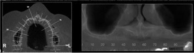
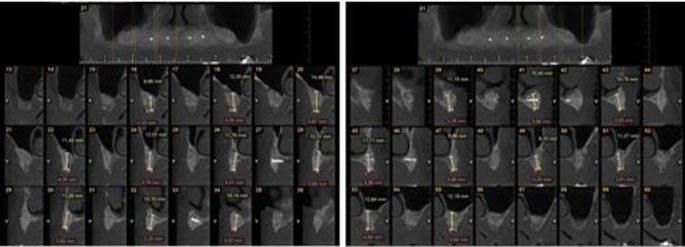
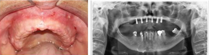
The 54-year-old leukoderma woman sought the Brazilian Dental Association (ABO) of Campo Grande, MS, Brazil, complaining that she had sought numerous oral rehabilitation centers without success, all of which retreated due to the great complexity of the case that presented severe maxillary resorption. In the initial consultation we were able to check on tomography regions of anterior premaxilla and maxillary sinus, where large regions of bone resorption were recorded (Figure 1).
Particulate biomaterial with the aid of platelet aggregate was planned for this case to regenerate the areas of the bilateral maxillary sinus and the entire anterior pre-maxillary region.
Biomaterial and dental implant
To reconstruct the area of bone loss found in this case, a biomaterial of alloplastic origin called Blue Bone (Curitiba, Brazil) was used. For the oral rehabilitation, dental implants, Cone Morse Avantt (CM), Systhex (Curitiba, Brazil) were selected.
Surgery and prosthetic stage
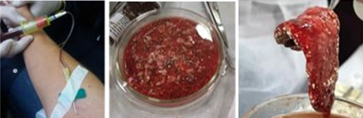
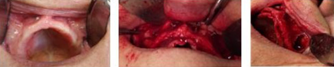
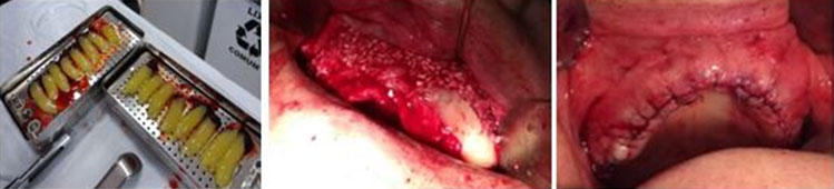
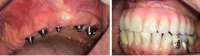
In order to collect a significant amount of blood for the preparation of the platelet aggregate, the patient was instructed to drink 3–4 liters of water per day, not to eat fried food or fats and to give preference to white meat for seven days. On the day of surgery, 28 tubes of the patient’s blood (using the Shokrum Dohan Ehrenfest DM) were collected to make the platelet aggregate and make the L-PRF membranes (Figure 2). The patient received antibiotics: 2 g amoxicillin (4 × 500 mg capsules) 1 hour after surgery and Clavulin 875 mg every 12 hours for 14 days and dipyrone (1 g tablet) for six days. A wash of 0.12% chlorhexidine solution for 1 minute was performed before the operation. Local anesthesia (2% lidocaine mepivacaine with adrenaline 1:100,000) was administered. Initially, a supra crystal incision was made from tuber to tuber with total displacement. Then, the left and right maxillary sinus were lifted, and tent screws were installed in the premaxilla region to better support the forces of the tissues on top of the graft (Figure 3). The right and left maxillary sinus and premaxilla were filled with 6 grams of nano-biomaterial (Blue Bone, Curitiba, Brazil) and, finally, the entire grafted area was covered with the L-PRF membranes (1300 rpm), and the sutures were performed (Figure 4). The patient was not using the total prosthesis for the first two months. Six months after surgery, a new tomography was performed, where we were able to analyze the extraordinary bone regeneration achieved in all grafted regions, achieving bone gain over tent screws (Figure 5). Six Cone morse Avantt model dental implants, 3.5 × 11 mm, from (Systhex, Curitiba, Brazil) (Figure 6) were planned. A six-month period was given for the osseo-integration, then the mini pillars were placed, and the case was rehabilitated with a Branemark resin protocol prosthesis (Figure 7).
DISCUSSION
Maximiano et al. [10], in a prospective randomized clinical study in humans, a total of 49 maxillary sinus floor lifts that were performed in 49 different patients were analyzed and divided into two groups: a control group (without graft, 25 patients) and a test group (with graft, 24 patients). The conclusion was that maxillary sinus elevation and implant placement without grafting is a successful treatment with few complications. But these statements oppose what the vast literature states, that the elevation and grafting of the maxillary sinus is a favorable and predictable procedure [11],[12],[13], based on the results presented in this study.
Bone augmentation techniques are not fully predictable and are not always able to guarantee the expected result, especially in the anterior region of the atrophic maxilla. Complications and failures can often occur, and this possibility should always be explained clearly to those patients with high demands and aesthetics [14]. However, other authors [15],[16] claim that proper selection of the implant to avoid oversized implants; careful handling and low soft tissue trauma and placement of the implant in an appropriate position using a periodontal probe or a prefabricated surgical guide; use of the appropriate surgical technique, such as guided bone regeneration with barrier membranes and appropriate bone grafts and or bone substitutes; and the precise closure of the wound establishes a favorable condition for a positive outcome of the case, just as the clinical case in this study presented was performed.
In addition, a meta-analysis that gathered a collection of randomized studies, where survival rate, bone neo-formation, contact between neo-formed bone and bone substitute with use and without the use of PRF were analyzed, showed that there was no statistically significant difference between the groups studied and the rate of implant loss was also not relevant, concluding that the current evidence supports the need to add PRF to the bone graft in the increase of the maxillary sinus and other areas are Limited’s [17]. One of the limitations of this study was not presenting a histological analysis, but in clinical and radiographic analyses they showed results that were extremely satisfactory and did not present loss of dental implants.
Oral rehabilitation in patients with protocol-style prostheses has been consolidated in the literature due to its positive clinical response in the short and long term, leading to quality of life for several patients [18],[19], but the available literature proves that a large percentage of these patients require rehabilitation with dental implants and need to associate the use of biomaterials for total resolution of the case [20],[21],[22]. In the study presented we can consolidate these statements by showing fantastic bone regeneration with a fixed prosthesis on implant in the Branemark protocol model.
CONCLUSION
Through the presented case report it is possible to conclude that cases of maxilla with large bone resorptions can be solved using surgical techniques with particulate nano-biomaterials associated with platelet aggregates, achieving predictable results in the short and long term.
REFERENCES
1.
da Silva Brum I, de Carvalho JJ, da Silva Pires JL, de Carvalho MAA, Dos Santos LBF, Elias CN. Nanosized hydroxyapatite and β-tricalcium phosphate composite: Physico-chemical, cytotoxicity, morphological properties and in vivo trial. Sci Rep 2019;9(1):19602. [CrossRef]
[Pubmed]

2.
da Silva Brum I, Frigo L, Lana Devita R, et al. Histomorphometric, immunohistochemical, ultrastructural characterization of a nano-hydroxyapatite/beta-tricalcium phosphate composite and a bone xenograft in sub-critical size bone defect in rat calvaria. Materials (Basel) 2020;13(20):4598. [CrossRef]
[Pubmed]
3.
Brum IS, Elias CN, de Carvalho JJ, Pires JLS, Pereira MJS, de Biasi RS. Properties of a bovine collagen type I membrane for guided bone regeneration applications. e-Polymers 2021;21(1):210–21. [CrossRef]
4.
Mangano C, Giuliani A, De Tullio I, Raspanti M, Piattelli A, Iezzi G. Case report: Histological and histomorphometrical results of a 3-D printed biphasic calcium phosphate ceramic 7 years after insertion in a human maxillary alveolar ridge. Front Bioeng Biotechnol 2021;9:614325. [CrossRef]
[Pubmed]
5.
Asa’ad F, Pagni G, Pilipchuk SP, Giannì AB, Giannobile WV, Rasperini G. 3D-printed scaffolds and biomaterials: Review of alveolar bone augmentation and periodontal regeneration applications. Int J Dent 2016;2016:1239842. [CrossRef]
6.
da Silva Brum I, Frigo L, Goncalo Pinto Dos Santos P, Nelson Elias C, da Fonseca GAMD, Jose de Carvalho J. Performance of nano-hydroxyapatite/beta-tricalcium phosphate and xenogenic hydroxyapatite on bone regeneration in rat calvarial defects: Histomorphometric, immunohistochemical and ultrastructural analysis. Int J Nanomedicine 2021;16:3473–85. [CrossRef]
[Pubmed]
7.
La Monaca G, Iezzi G, Cristalli MP, Pranno N, Sfasciotti GL, Vozza I. Comparative histological and histomorphometric results of six biomaterials used in two-stage maxillary sinus augmentation model after 6-month healing. Biomed Res Int 2018;2018:9430989. [CrossRef]
[Pubmed]
8.
Azim AA, Wang HH, Serebro M. Selective retreatment and sinus lift: An alternative approach to surgically manage the palatal roots of maxillary molars. J Endod 2021;47(4):648–57. [CrossRef]
[Pubmed]
9.
Carosi P, Lorenzi C, Lio F, Laureti M, Ferrigno N, Arcuri C. Short implants (≤6mm) as an alternative treatment option to maxillary sinus lift. Int J Oral Maxillofac Surg 2021;S0901-5027(21)00055-2. [CrossRef]
[Pubmed]
10.
Maximiano Millán A, Bravo Álvarez R, Plana Montori M, et al. Assessment of the simultaneous use of biomaterials in transalveolar sinus floor elevation: Prospective randomized clinical trial in humans. Int J Environ Res Public Health 2020;17(6):1888. [CrossRef]
[Pubmed]
11.
Zhao X, Gao W, Liu F. Clinical evaluation of modified transalveolar sinus floor elevation and osteotome sinus floor elevation in posterior maxillae: Study protocol for a randomized controlled trial. Trials 2018;19(1):489. [CrossRef]
[Pubmed]
12.
Guerrero JS, Al-Jandan BA. Lateral wall sinus floor elevation for implant placement: Revisiting fundamentals and the surgical technique. J Calif Dent Assoc 2013;41(3):185–7, 190–5. [CrossRef]
[Pubmed]
13.
Yu SJ, Lee YH, Lin CP, Wu AYJ. Computed tomographic analysis of maxillary sinus anatomy relevant to sinus lift procedures in edentulous ridges in Taiwanese patients. J Periodontal Implant Sci 2019;49(4):237–47. [CrossRef]
[Pubmed]
14.
Checchi V, Gasparro R, Pistilli R, Canullo L, Felice P. Clinical classification of bone augmentation procedure failures in the atrophic anterior maxillae: Esthetic consequences and treatment options. Biomed Res Int 2019;2019:4386709. [CrossRef]
[Pubmed]
15.
Buser D, Martin W, Belser UC. Optimizing esthetics for implant restorations in the anterior maxilla: Anatomic and surgical considerations. Int J Oral Maxillofac Implants 2004;19 Suppl:43–61.
[Pubmed]
16.
Hyun YK, Lee CY, Keerthana S, et al. Horizontal alteration of anterior alveolar ridge after immediate implant placement: A retrospective cone beam computed tomography analysis. J Adv Prosthodont 2021;13(2):117–25. [CrossRef]
[Pubmed]
17.
Dohan Ehrenfest DM, Doglioli P, de Peppo GM, Del Corso M, Charrier JB. Choukroun’s platelet-rich fibrin (PRF) stimulates in vitro proliferation and differentiation of human oral bone mesenchymal stem cell in a dose-dependent way. Arch Oral Biol 2010;55(3):185–94. [CrossRef]
[Pubmed]
18.
Liu R, Yan M, Chen S, Huang W, Wu D, Chen J. Effectiveness of platelet-rich fibrin as an adjunctive material to bone graft in maxillary sinus augmentation: A meta-analysis of randomized controlled trails. Biomed Res Int 2019;2019:7267062. [CrossRef]
[Pubmed]
19.
Liu CS. Periodontal prosthesis in contemporary dentistry. Kaohsiung J Med Sci 2018;34(4):194–201. [CrossRef]
[Pubmed]
20.
Maló P, Rangert B, Nobre M. “All-on-Four” immediate-function concept with Brånemark System implants for completely edentulous mandibles: A retrospective clinical study. Clin Implant Dent Relat Res 2003;5 Suppl 1:2–9. [CrossRef]
[Pubmed]
21.
Göthberg C, Gröndahl K, Omar O, Thomsen P, Slotte C. Bone and soft tissue outcomes, risk factors, and complications of implant-supported prostheses: 5-Years RCT with different abutment types and loading protocols. Clin Implant Dent Relat Res 2018;20(3):313-21. [CrossRef]
[Pubmed]
22.
deF Silva L, de Lima VN, Faverani LP, de Mendonça MR, Okamoto R, Pellizzer EP. Maxillary sinus lift surgery-with or without graft material? A systematic review. Int J Oral Maxillofac Surg 2016;45(12):1570–6. [CrossRef]
[Pubmed]
SUPPORTING INFORMATION
Acknowledgments
This research received financial support from the Brazilian Research Agency CNPq, FAPERJ, UERJ, Capes, and Lubt.
Author ContributionsGustavo Helder Vinholi - Conception of the work, Design of the work, Acquisition of data, Analysis of data, Drafting the work, Revising the work critically for important intellectual content, Final approval of the version to be published, Agree to be accountable for all aspects of the work in ensuring that questions related to the accuracy or integrity of any part of the work are appropriately investigated and resolved.
Igor da Silva Brum - Conception of the work, Design of the work, Acquisition of data, Analysis of data, Drafting the work, Revising the work critically for important intellectual content, Final approval of the version to be published, Agree to be accountable for all aspects of the work in ensuring that questions related to the accuracy or integrity of any part of the work are appropriately investigated and resolved.
Renan Lana Devita - Conception of the work, Design of the work, Acquisition of data, Analysis of data, Drafting the work, Revising the work critically for important intellectual content, Final approval of the version to be published, Agree to be accountable for all aspects of the work in ensuring that questions related to the accuracy or integrity of any part of the work are appropriately investigated and resolved.
João Carlos Lopes Amorim - Conception of the work, Design of the work, Acquisition of data, Analysis of data, Drafting the work, Revising the work critically for important intellectual content, Final approval of the version to be published, Agree to be accountable for all aspects of the work in ensuring that questions related to the accuracy or integrity of any part of the work are appropriately investigated and resolved.
Marco Antônio Alencar de Carvalho - Conception of the work, Design of the work, Acquisition of data, Analysis of data, Drafting the work, Revising the work critically for important intellectual content, Final approval of the version to be published, Agree to be accountable for all aspects of the work in ensuring that questions related to the accuracy or integrity of any part of the work are appropriately investigated and resolved.
Jorge José de Carvalho - Conception of the work, Design of the work, Acquisition of data, Analysis of data, Drafting the work, Revising the work critically for important intellectual content, Final approval of the version to be published, Agree to be accountable for all aspects of the work in ensuring that questions related to the accuracy or integrity of any part of the work are appropriately investigated and resolved.
Guaranter of SubmissionThe corresponding author is the guarantor of submission.
Source of SupportNone
Consent StatementWritten informed consent was obtained from the patient for publication of this article.
Data AvailabilityAll relevant data are within the paper and its Supporting Information files.
Conflict of InterestAuthors declare no conflict of interest.
Copyright© 2021 Gustavo Helder Vinholi et al. This article is distributed under the terms of Creative Commons Attribution License which permits unrestricted use, distribution and reproduction in any medium provided the original author(s) and original publisher are properly credited. Please see the copyright policy on the journal website for more information.


