 |
Original Article
The interrelationship of arbitrary surface inter-condylar distance, freeway space, rest vertical dimension and occlusal vertical dimension
1 Conservative Dentistry Unit, Department of Restorative Dentistry, Faculty of Dentistry, Lagos State University College of Medicine Ikeja, Nigeria
2 Prosthetic Dentistry Unit, Department of Restorative Dentistry, Faculty of Dentistry, Lagos State University College of Medicine Ikeja, Nigeria
Address correspondence to:
Adenike Ololade Awotile
Conservative Dentistry Unit, Department of Restorative Dentistry, Faculty of Dentistry, Lagos State University College of Medicine Ikeja,
Nigeria
Message to Corresponding Author
Article ID: 100038D01AO2019
Access full text article on other devices

Access PDF of article on other devices


How to cite this article
Awotile AO, Loto AO, Adenuga-Taiwo OA. The interrelationship of arbitrary surface inter-condylar distance, freeway space, rest vertical dimension and occlusal vertical dimension. Edorium J Dent 2019;6:100038D01AO2019.ABSTRACT
Aims: To find the correlation between arbitrary surface intercondylar distance on one hand and freeway space, rest vertical dimension, occlusal vertical dimension on the other hand.
Methods: A cross sectional study of randomly selected individuals between 18–56 years old, in a tertiary hospital in Ikeja from September- October 2017. Participants were examined and measurements, arbitrary surface intercondylar distance, rest vertical dimension,and occlusal vertical dimension were taken. Data collected were subjected to statistical analysis using SPSS version 20. Level of significance was set at p≤0.05.
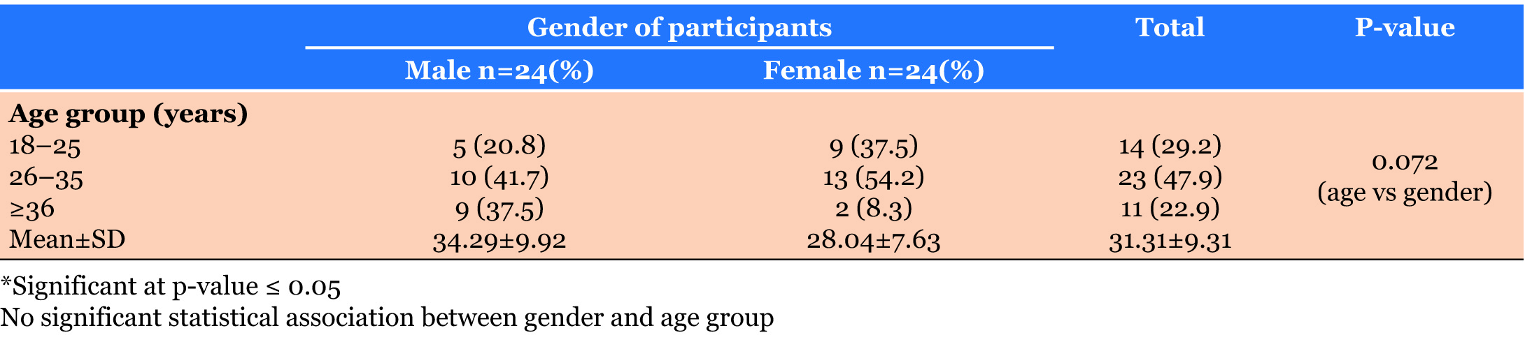
Results: Of the 48 participants involved in this study, 50% were males. Majority (47.9%) were between 26–35 years old with a mean age of 31 years. Participants in age group 18–25 years and ≥36 years had similar freeway space (FWS) which were 3.43±1.01 mm and 3.45±0.82 mm respectively while 26–35 years old had 3.13±0.87 mm. Mean FWS was 3.29 mm. There was no significant statistical difference in FWS based on gender and age. Spearmans correlation coefficient (R2) analysis revealed a positive correlation of intercondylar distance with freeway space, rest vertical dimension and occlusal vertical dimension. Regression equations were formulated and utilized to calculate concerned parameters.
Conclusion: There is correlation between inter condylar distance on one hand and freeway space, rest vertical dimension and occlusal vertical dimension on the other hand. Therefore a regression equation becomes a simple and rapid method of calculating each parameter.
Keywords: Freeway space, Intercondylar distance, Rest vertical dimension, Occlusal vertical dimension
INTRODUCTION
Freeway space (FWS) otherwise known as interocclusal clearance or inter-occlusal distance or occlusal rest space or inter-occlusal space is the distance between the occlusal and incisal surfaces of upper and lower antagonistic teeth when the mandible is in physiologic rest position or postural position [1]. It is the difference between rest vertical dimension (RVD) and occlusal vertical dimension (OVD). Occlusal vertical dimension (OVD) is the height of the lower part of the face measured from two reference points when the maxillary and mandibular teeth (dental arches) are in centric occlusion and is influenced by various factors while rest vertical dimension (RVD) is the height of the lower part of the face measured from identified two reference points when the mandible is in physiologic rest position.
The most commonly used reference points for measuring, OVD and RVD are subnasale and menton; and tip of the nose and gnathion. These three parameters (FWS, OVD & RVD) are separate but inter-related physiologic parameters. They are of great importance in diagnosis, treatment planning as well as treatment procedures’ evaluation in operative dentistry, prosthodontics, orthodontics and maxillo-facial surgery. In fully edentulous people, there are no teeth to ensure mutual contact between upper and lower dental arches. Therefore, jaw relationship when fabricating dentures, should be re-established with artificial dental arches in the proper vertical and horizontal relationships. This is one of the most complex procedures in prosthodontics [2].
The role of FWS in re-constructive restorative dentistry cannot be over emphasized; and the importance of OVD and RVD in the determination of FWS cannot be under-estimated because FWS is derived from RVD and OVD. Therefore, the accuracy of FWS depends on the degree of accuracy of the determination of RVD and OVD.
OVD in a dentate patient is relatively constant throughout life, even in the face of severe generalized attrition or localized attrition involving only the anterior teeth. This relative constancy is maintained by compensatory growth of the alveolar process which tries to restore the OVD to its initial status. Complete loss of teeth or loss of posterior teeth results in reduced OVD because OVD depends on the height of the ramus and the clinical heights of the posterior teeth. RVD is a variable parameter which cannot be determined precisely by all known methods. It is influenced by various factors such as head posture, stress, fatigue, loss of teeth, age, health status, bruxism, methods of its determination as well as the skillfulness of the dentists during RVD’s determination.
Various methods have been reportedly used in the determination of RVD and OVD in both dentate and edentulous people by different authors. These methods include pre-extraction records, photographs, swallowing method, phonetic method, facial anthropometric measurements, transcutaneous electrical nerve stimulations (TENS) and so on. It is generally agreed that none of these methods can accurately and reliably determine RVD and OVD. Consequently, incorrect determination of RVD and/or OVD will result in incorrect determination of FWS. FWS can vary within an individual and from individual to individual. It may be affected by factors such as head posture, age, mental state, fatigue, some medications etc. Adaptive FWS is defined as the inter-occlusal space that exists when patient is instructed to voluntarily allow the lower jaw to relax [3] while the true FWS is defined as the measured inter-occlusal space when the lower jaw has acquired position of least muscular activity after TENS by the myomonitor [3],[4].
The average FWS normally employed in prosthodontics procedure is 2–4 mm. However, FWS like RVD & OVD, is dynamic and depends on OVD and RVD. It is essential for speech, efficient mastication, aesthetics and comfort. Consequently, reduced or increased FWS will impact negatively on speech, mastication, facial aesthetics and comfort. In view of the aforementioned importance of FWS, dentists are constantly reviewing and seeking more effective, efficient and accurate methods or ways of determining FWS, OVD and RVD by comparing facial anthropometric measurements using proportions or ratios. One of the facial anthropometric measurements of great interest is the bi-condylar distance which is defined as the distance between the left and right condyles measured from defined or specified reference points. The following possible combinations of reference points can be used: skin surfaces located just in front of the two tragi to mark the head of the two mandibular condyles, lateral surfaces (poles) of the two condyles, lateral surface of one condyle and medial surface of the other condyle, the medial surfaces of the two condyles, the lateral surface of one condyle and the mid-point of the medio-lateral dimension of the other condyle; and the mid-point of the medio-lateral width of the two condyle [5]. Therefore, in the determination of the inter-condylar distance, the researcher must clearly state the reference points that were used in order to avoid confusion.
Bi-condylar distance (intercondylar distance) has been widely studied by various researchers who reported variations according to gender, age, race and facial type [6],[7],[8]. This parameter is one of the most stable and constant facial anthropometric measurements [6],[9],[10]. The relative constancy of this parameter has enabled many clinicians, researchers and anthropologists to associate it with other facial anthropometric parameters for the purpose of establishing definitive proportions or ratios between it and other facial measurements. For examples, intercondylar distance has been correlated with lower anterior facial height (LAFH), upper anterior facial height (UAFH), sum of the mesio-distal dimensions of the six anterior teeth in mandible and maxilla, and the sum of the mesio-distal widths of four upper incisors [6],[7],[8],[9],[10].
The need to search for more accurate methods for the determination of the aforementioned facial parameters cannot be overemphasized. Consequently, the objectives of this study were:
- To determine whether there is any correlation between bi-condylar distance, OVD and RVD on one hand and FWS on the other hand;
- To formulate regression equations, based on correlation co-efficient, for calculating bicondylar distance, OVD, RVD and FWS;
- To compare the results of this study with previous local and international studies.
MATERIALS AND METHODS
This is a descriptive cross-sectional study which was carried out on forty-eight (48) randomly selected patients who attended the oral diagnosis clinic at the Lagos State University Teaching Hospital, Ikeja, Lagos, Nigeria from September to October 2017. There were 24 males and 24 females with age range of 18–56 years and combined mean age of 31years.
The inclusion criteria for the participants included:
- Full upper and lower dentitions with Angle’s class I molar and canine relationships
- No history of TMJ disorders (symptoms and signs)
- Absence of restorations, bridges and dentures
- No history of jaw fracture and surgical operation
- No facial asymmetry
- Willingness to participate in the study and sign the consent form after the procedures had been carefully explained to each participant.
The exclusion criteria included in following:
- Incomplete upper and lower dentitions
- Presence of malocclusion 3. History of temporomandibular joint disorders
- Presence of restorations, bridges and dentures
- History of jaw fracture and operation
- Facial asymmetry
- Patient whose age is below 18 years or above 45 years.
Ethical approval for the study protocol was obtained from the Health Research Ethical Committee of LASUTH. Written informed consent was also obtained from patients and they were assured of confidentiality.
The following anthropometric parameters were measured as follows:
Determination of bi-condylar distance
Each participant was seated upright, comfortably on the dental chair, looking straight forward with the head in erect position. The left and right condyles were palpated just in front of the tragi and their positions were noted. The condylar rods of the arbitrary face bow were made to touch the marked surface points of the condylar head and the distance between the left and right condyles was read from a metre rule. Two measurements were made by a single investigator who was previously calibrated for the purpose of accuracy and reliability and having intraexaminer coefficient of variation of 0.99. The average value of the two measurements for each participant was determined by dividing sum of the two measurements by two. The values obtained for the participants were recorded in a form designed for this purpose.
Determination of OVD, RVD and FWS
Each participant was seated upright, comfortably on the dental chair with the head in erect position and the occlusal plane was made parallel to the floor. The subject was instructed to bring the upper and lower posterior teeth into close contact and the distance between the subnasale and menton was measured twice using Alma gauge and the values were read on this Alma gauge to the nearest 0.1 mm. The two measurements were summed up and then divided by two to obtain average value for each participant. This value represented the OVD of each participant.
To obtain the RVD, each participant, while maintaining the same sitting position in a relaxed mood, was also instructed to relax his lower jaw and to allow the upper and lower lips just to contact slightly. The distance between the subnasale and menton was measured twice using Alma gauge; and the average value of the two measurements was determined to represent the RVD for each participant. The difference between mean RVD and mean OVD, for each participant, was taken as the freeway space (FWS) of that participant.
To obtain the FWS for each participant, the average value of OVD was subtracted from corresponding mean value of RVD for each subject.
All the obtained values for RVD, OVD and FWS were recorded in the form designed for this purpose. The data obtained from the various measurements in this study were subjected to statistical analysis using SPSS version 20. Descriptive statistics such as frequency, mean and standard deviation were used for the purpose of formulating tables and figures. Student’s test was used to compare the mean differences between two variables and Spearman’s correlation coefficient was used to determine any correlation or association among the various variables. Level of significance was set as p<0.05. Simple regression analysis was used to formulate regression equation for calculating freeway space.
Sample size determination
N = 16 × (SD)2 ÷ W2
N = Sample size;
SD= Standard Deviation
W = Width of maximum margin of error
SD referenced in study (4) = ±1.6
W = 1
N = 16×(1.6×1.6) ÷ 1=40.96 (approx. 41)
Plus 20% attrition = 8.192 = 8
Therefore sample size = 41+8 = 49
RESULTS
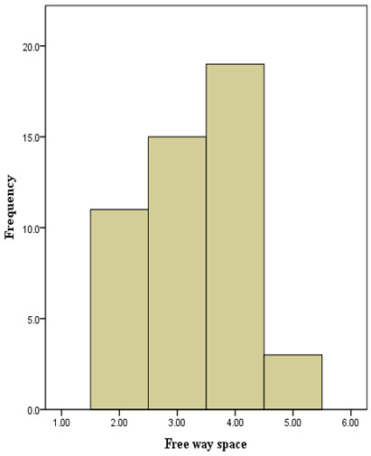
There were 48 participants involved in this study. The majority (47.9%) of the participants were between 26–35 years old with a mean age of 31 years. There was no significant statistical association between gender and age group (Table 1). Most of the participants had a FWS of about 4 mm while quite a few had 5 mm. The mean value of FWS was 3.29 mm (Figure 1).
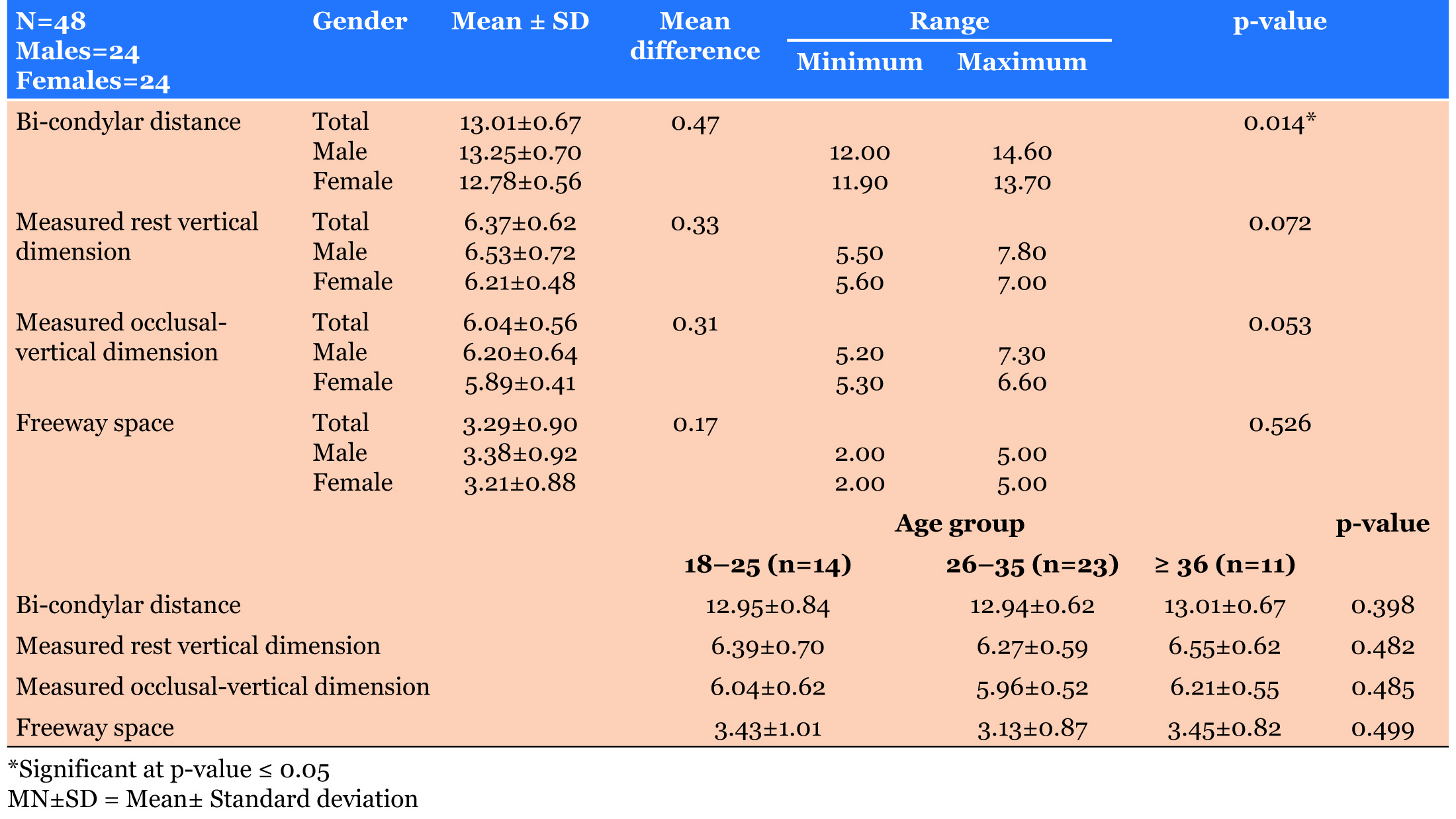
The participants in the age groups 18–25 years and >36 years had similar mean values of FWS 3.43 ±1.01 mm and 3.45±0.82 mm respectively. The mean FWS of 26–35 years age group was slightly smaller (3.13±0.87 mm) than the other age groups. However, there was no statistically significant difference in the value of FWS based on age group (Table 2). Males had an average mean value of 3.38±0.92 mm FWS while females had 3.21±0.88 mm FWS with a p-value of 0.526; hence, no significant statistical difference was seen in FWS based on with a p-value of 0.526; hence, no significant statistical difference was seen in FWS based on gender (Table 2).
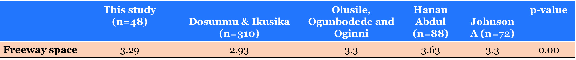
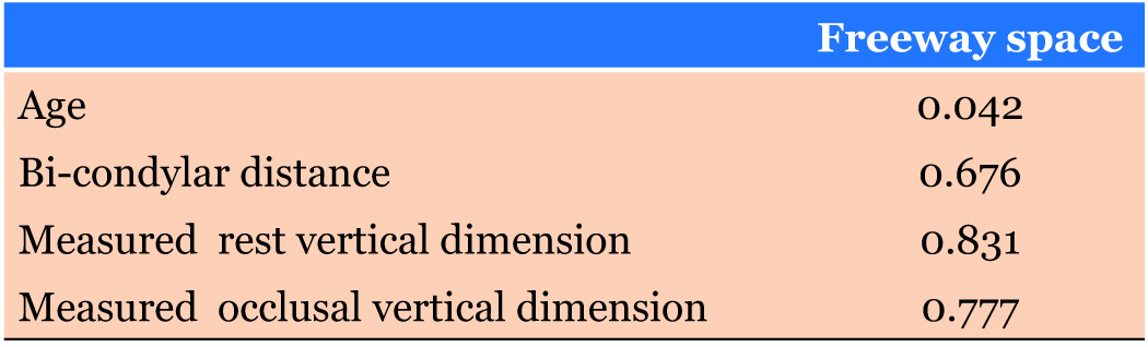
Mean comparison of freeway space of this study with an international study using Turkey post hoc analysis showed a significant difference [11] while no difference was deduced when compared to local studies [12],[13],[14] (Table 3). Spearman’s correlation coefficient analysis revealed a positive correlation of freeway space with rest vertical dimension, intercondylar distance and occlusal vertical dimension whereas it showed a weak positive correlation with age (Table 4).
Based on the obtained correlation coefficient analysis (R2) values, the model has a high predictive ability to calculate the freeway space using measured occlusal vertical dimension, rest vertical dimension and bicondylar distance (Table 4).
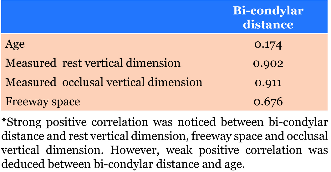
Spearman’s correlation coefficient analysis also revealed a positive correlation of bi-condylar distance with rest vertical dimension, freeway space and occlusal vertical dimension whereas it showed a weak positive correlation with age (Table 5). Based on the obtained correlation coefficient analysis (R2) values, the model has a strong ability to predict the bi-condylar distance using freeway space, rest vertical dimension and occlusal vertical dimension.
Simple regression equations were formulated as follows:
Predicting freeway space using measured rest vertical dimension
Freeway space = -4.279+1.188 (measured rest vertical dimension)
R2=0.831
Predicting freeway space using measured occlusal vertical dimension
Freeway space = -4.300+1.157 (measured occlusal vertical dimension)
R2=0.777
Predicting freeway space using measured bi-condylar distance
Freeway space = -8.437+0.901 (measured bicondylar distance)
R2=0.676
Predicting bi-condylar distance using measured rest vertical dimension
Bi-condylar distance= 6.845+0.969 (measured rest vertical dimension)
R2=0.902
Predicting bi-condylar distance using measured occlusal vertical dimension
Bi-condylar distance = 6.341+1.105 (measured occlusal vertical dimension)
R2= 0.911
Predicting bi-condylar distance using freeway space
Bi-condylar distance=11.349+0.507 (freeway space)
R2= 0.676
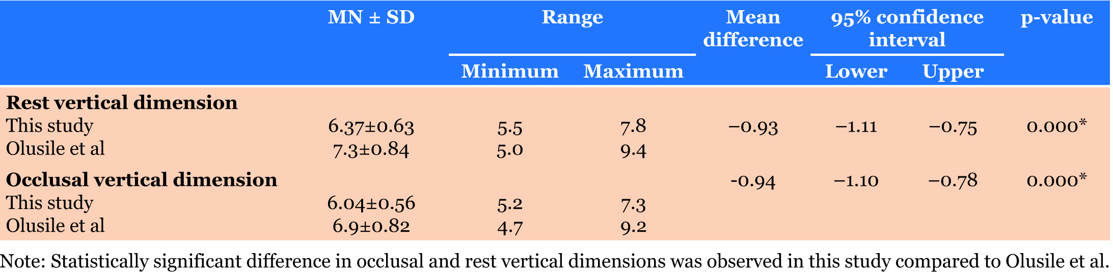
Table 6 shows comparison of mean values of RVD and OVD in this study with another study [13] which was found to have higher mean values for RVD and OVD relative to our study with the mean difference statistically significant (p-value = 0.000).
DISCUSSION
Freeway space can be obtained by subtracting the value of OVD from the value of RVD. It is generally accepted that some oral and facial anatomical sites remain relatively stable throughout life [15] hence the utilization of such landmarks (width of the mouth, inter-alar width, bi-zygomatic width, and inter-pupillary distance) in the determination of OVD and RVD. However, these landmarks should be used as combined methods rather than being used individually so as to reduce errors arising from racial and gender differences [9].
This present study showed an average freeway space of 3.29 mm. This was found to be similar to some studies [12],[13],[14] but there was a statistical significant difference (p-value =0.00) between this study and that among the Arabs [11], which was 3.63 mm using Turkey post hoc analysis. The difference between this study and latter [11] might be due to racial difference since our study, and those of African studies [12],[13],[14] were carried out among Negroes while the rest took place among Arabs [11]. The mean value of FWS obtained in this present study also falls within the ranges that were reported by other researchers [4],[16],[17],[18].
In this study, the average values of freeway space (FWS) for the three classified groups (18–25 years, 26–35 years and 36 years and above) were 3.43±1.01 mm, 3.13±0.87 mm and 3.45±0.82 mm respectively. However, it was found that the mean values of FWS for the age groups 18–25 years and 36 years and above were similar (Table 2) while the average value of FWS for the age group 26–35 years was slightly smaller than the other two groups. Nonetheless, the mean difference was not statistically significant (p-value = 0.499). The slightly smaller mean value of FWS for age group 26–35 years could be attributed to larger number of subjects (23) in that age group compared to the other two age groups (18–25 years had 14 participants and 36 years and above had 11 participants). The statistically insignificant difference in the average values of FWS based on age groups suggests that age might not have any significant effect on FWS.
Comparison of mean values of bicondylar distance based on age groups revealed average values of 12.95+0.62 cm, 12.94±0.62 cm and 13.01+0.67 cm for groups of 18–25 years, 26–35 years and 36 years and above respectively. It was found that no statistical significant difference existed among the three groups (p-value=0.398). The statistically insignificant difference in the average values of bi-condylar distance among the three age groups could be attributed to early establishment or attainment of the major proportion of the size of the intercondylar distances during pubertal period of the growth of mandible. Postpubertal increase in the size of the mandible is minimal. Therefore, it has no significant effect on the size of the intercondylar distances among the different age groups of the study population [19],[20],[21],[22].
This study also showed sexual dimorphism with respect to intercondylar distance as it had been observed by several previous studies [6],[7],[8]. In this study, the mean values for male and female were 13.25+0.70 cm and 12.78+0.56 cm respectively while the combined average value of bi-condylar distance for both sexes was 13.01+0.67 cm. These average values of surface bi-condylar distances closely correlate with the average values of arbitrary surface intercondylar distances obtained in their respective studies [6],[7]. Two types of intercondylar distance (arbitrary surface intercondylar distance and kinematic surface intercondylar distance) were determined for the study population [7]. The kinematic intercondylar distances and arbitrary intercondylar distances were obtained using Kinematic face bow and arbitrary face bow, for measuring intercondylar distance, respectively.
However, the mean values of bicondylar distances obtained in our study could not be directly compared with that of a particular study with a significantly differentmethodology. In terms of definition of reference points for measurement of intercondylar distance, the reference points for measurement in our study were surface landmarks that were marked on the skin surfaces of the condylar heads just in front of the two tragi while in the other study [8], the intercondylar distances were determined on posterior-anterior radiographs using lateral poles of the two condyles as the reference points for measuring the intercondylar distances. Therefore, in order to have a scientifically sound comparison between our study and this particular study [8], the 2.5 cm that represents the combined skin thickness dimensions over the two condyles must be deducted from our study. Consequently, the mean values of bicondylar distances for male and female, in our study, would be 10.75 cm (107.5 mm) and 10.28 cm (102.8 mm) respectively while the combined mean value for both sexes would be 10.51 cm (105.1 mm). Therefore, the average values of bi-condylar distances, 13.02 cm (130.2 mm) for male, 12.35 cm (123.5 mm) for female and 12.6 cm (126 mm) for both sexes, obtained [8] in their study were significantly greater than average values of bicondylar distances obtained in our study.
Bi-condylar distance (inter-condylar distance), a fairly static parameter throughout life, has been correlated and found to have a strong positive association with maxillary and mandibular inter-canine distance thereby aiding in the selection of maxillary and mandibular anterior six teeth in complete dentures [9],[10],[23]. From our study, we also found out that we could calculate freeway space from OVD, RVD and bi-condylar distance values using simple regression equations based on the correlation coefficient (R2) values. This mathematical model has a high predictive ability to calculate the freeway space using measured occlusal vertical dimension, rest vertical dimension and bi-condylar distance as shown below:
In this study, bi-condylar distance was also correlated with rest vertical dimension, occlusal vertical dimension and freeway spaceusing Spearman’s correlation analysis; and it was found that intercondylar distance exhibited strong positive relationships with rest vertical dimension (R2 =0.902), occlusal vertical dimension (R2 =0.911) and freeway space (R2 = 0.676). Thus, the value of bi-condylar distance can be calculated from known or measured OVD, RVD and FWS using the formulated simple regression equations as given previously.
These regression equations are simple; and they constitute the quickest way of calculating OVD, RVD, FWS and bi-condylar distance. However, this mathematical model should be used in combination with other known techniques.
CONCLUSION
This study showed moderate correlation between bicondylar distance on one hand and OVD, RVD and FWS on the other hand. Regression equations were deduced for a quick calculation of freeway space, OVD, RVD and bi-condylar distance. The mean and range of freeway space values obtained in this study were also consistent with previous local and international studies. Further studies should be carried out to verify the authenticity of the simple regression equations obtained in this study.
REFERENCES
1.
Chou TM, Moore DJ, Young LJ Jr, Glaros AG, Chou JI. Occlusal vertical dimension in prosthodontics. Kaohsiung J Med Sci 1996;12(5):260–6.
[Pubmed]

2.
Guitierrez Da Venezia P. The evaluation of occlusal vertical dimension. J Dent Quebec 2003;40:241–3.
3.
Jankelson B, Radke JC. The myo-monitor: Its use and abuse (I). Quintessence Int Dent Dig 1978;9(2):47–52.
[Pubmed]
4.
Konchak PA, Thomas NR, Lanigan DT, Devon R. Vertical dimension and freeway space: A kinesiographic study. Angle Orthod 1987;57(2):145–54.
[Pubmed]
5.
Woelfel JB, Scheid RC, Weiss G. Woelfel's Dental Anatomy: Its Relevance to Dentistry. 8ed. Philadelphia: Lippincott Williams & Wilkins; 2012. p. 31–2.
6.
Qamar K, Shaikh IA, Naeem S. Relationship of the inter-condylar width with mandibular inter-canine width. J Ayub Med Coll Abbottabad 2013;25(1–2):191–3.
[Pubmed]
7.
Keshvad A, Winstanley RB, Hooshmand T. Intercondylar width as guide to setting up complete denture teeth. J Oral Rehabil 2000;27(3):217–26. [CrossRef]
[Pubmed]
8.
Lazić B, Tepavcević B, Keros J, Komar D, Stanicić T, Azinović Z. Intercondylar distances of the human temporomandibular joints. Coll Antropol 2006;30(1):37–41.
[Pubmed]
9.
Dahri WM, Butt AM, Ahmed B. Relationship of intercondylar distance with intercanine distance in dental students. J Pak Dent Assoc 2012;21(3):141–4.
10.
Debnath N, Gupta R, Meenakshi A, Kumar S, Hota S, Rawat P. Relationship of inter-condylar distance with inter-dental distance of maxillary arch and occlusal vertical dimension: A clinical anthropometric study. J Clin Diagn Res 2014;8(12):ZC39–43. [CrossRef]
[Pubmed]
11.
Abdul-Rahman H. Analysis of the relation between lip length, free way space, closest speaking space, arch size concerning palatal-depth relativity. MDJ 2009;6(2):152–8.
12.
Johnson A, Wildgoose DG, Wood DJ. The determination of freeway space using two different methods. J Oral Rehabil 2002;29(10):1010–3.
[Pubmed]
13.
Olusile AO, Ogunbodede EO, Oginni AO. Prosthetic parameters among dental patients in Ile-Ife, Nigeria. Niger Postgrad Med J 2003;10(2):88–91.
[Pubmed]
14.
Dosunmu OO, Ikusika OF. An assessment of interocclusal space in a dentate Nigerian population. Niger Postgrad Med J 2013;20(4):315–8.
[Pubmed]
15.
Valette C, Albouy JC, Ravon P. Determination of the vertical dimension of occlusion. [Article in French]. Cah Prothese 1989;(65):90–102.
[Pubmed]
16.
Nielsen IL, Marcel T, Chun D, Miller AJ. Patterns of mandibular movements in subjects with craniomandibular disorders. J Prosthet Dent 1990;63(2):202–17. [CrossRef]
[Pubmed]
17.
Ferrario VF, Sforza C, Miani A, D'Addona A, Tartaglia G. Statistical evaluation of some mandibular reference positions in normal young people. Int J Prosthodont 1992;5(2):158–65.
[Pubmed]
18.
Martín C, Alarcón JA, Palma JC. Kinesiographic study of the mandible in young patients with unilateral posterior crossbite. Am J Orthod Dentofacial Orthop 2000;118(5):541–8. [CrossRef]
[Pubmed]
19.
Goldstein M. Changes in dimensions and form of the face and head with age. Am J Phys Anthropol 1936;22(1):37–89. [CrossRef]
20.
Tofani MI. Mandibular growth at puberty. Am J Orthod 1972;62(2):176–95.
[Pubmed]
21.
Gomes AS, Lima EM. Mandibular growth during adolescence. Angle Orthod 2006;76(5):786–“9.
[Pubmed]
22.
Mitani H, Sato K. Comparison of mandibular growth with other variables during puberty. Angle Orthod 1992;62(3):217–22.
[Pubmed]
23.
Shaikh IH, Qamar K, Naeem S. Relationship of the intercondylar distance with maxillary intercanine distance. Pak Oral Dental J 2011;31:470–3.
SUPPORTING INFORMATION
Author Contributions
Adenike Ololade Awotile - Conception of the work, Design of the work, Acquisition of data, Analysis of data, Drafting the work, Revising the work critically for important intellectual content, Final approval of the version to be published, Agree to be accountable for all aspects of the work in ensuring that questions related to the accuracy or integrity of any part of the work are appropriately investigated and resolved.
Adulphus Odogun Loto - Conception of the work, Design of the work, Acquisition of data, Analysis of data, Drafting the work, Revising the work critically for important intellectual content, Final approval of the version to be published, Agree to be accountable for all aspects of the work in ensuring that questions related to the accuracy or integrity of any part of the work are appropriately investigated and resolved.
Olugbenga Adetokunbo Adenuga-Taiwo - Conception of the work, Design of the work, Acquisition of data, Analysis of data, Drafting the work, Revising the work critically for important intellectual content, Final approval of the version to be published, Agree to be accountable for all aspects of the work in ensuring that questions related to the accuracy or integrity of any part of the work are appropriately investigated and resolved.
Guaranter of SubmissionThe corresponding author is the guarantor of submission.
Source of SupportNone
Consent StatementWritten informed consent was obtained from the patient for publication of this article.
Data AvailabilityAll relevant data are within the paper and its Supporting Information files.
Conflict of InterestAuthors declare no conflict of interest.
Copyright© 2019 Awotile Adenike Ololade et al. This article is distributed under the terms of Creative Commons Attribution License which permits unrestricted use, distribution and reproduction in any medium provided the original author(s) and original publisher are properly credited. Please see the copyright policy on the journal website for more information.


