|
Research Article
Diode laser versus conventional surgery for ankyloglossia: A case series comparison
1 Professor, Department of Periodontics, Iguaçu University, Nova Iguaçu, Rio de Janeiro, Brazil
2 Professor, Department of Oral Surgery, Iguaçu University, Nova Iguaçu, Rio de Janeiro, Brazil
3 Professor, Department of Pediatric Dentistry, Iguaçu University, Nova Iguaçu, Rio de Janeiro, Brazil
Address correspondence to:
Carlos Henrique Sardenberg Pereira
Rua Conde de Bomfim n232, apt 908, CEP 20.520-054, Tijuca, Rio de Janeiro, Rio de Janeiro,
Brazil
Message to Corresponding Author
Article ID: 100047D01CP2025
Access full text article on other devices

Access PDF of article on other devices


How to cite this article
Pereira CHS, Magro OO, Nunes VHT, de Uzeda MJPG, Brum SC. Diode laser versus conventional surgery for ankyloglossia: A case series comparison. Edorium J Dent 2025;12(2):1–8.ABSTRACT
Ankyloglossia is a condition in which the tongue cannot protrude beyond the lower incisor teeth due to the presence of abnormal shortened lingual frenulum. The present study evaluated post-surgical discomfort of and healing aspects in two different groups, including three patients diagnosed with ankyloglossia each. Individuals in group A underwent regular surgical procedure, while for patients in group B were treated by a diode laser device (Laser Tw Surgical, MMoptics, Brazil). The results demonstrate patients undergoing laser-assisted frenectomy reported that pain is significantly reduced when compared to patients undergoing conventional surgical frenectomy, both immediately after surgery and one week after the first week, also, better signs of wound healing was observed in group B. In addition, some advantages were reported by the operator during the procedure in laser-assisted group: absence of bleeding and, consequently, a clear operative field; no need to use sutures; no need to take painkillers or antibiotics after surgery and a shorter operation duration needed. Within the limits of the present study, it seems possible to state that laser frenectomy has a number of advantages over the conventional surgical method.
Keywords: Ankyloglossia, Frenectomy, Laser, Surgery
INTRODUCTION
Ankyloglossia (tongue-tie) is a congenital oral anomaly characterized by a shortened and tightened lingual frenulum that affects tongue movements and functions such as sucking, speaking, and eating [1]. This condition can be found in newborns and children, as well as adolescents and adults [2]. Tongue-tie in nursling infants is related to difficulty breastfeeding (choking, spitting up) delayed speech development, speech complaints, and behavioral problems [3],[4].
Lingual frenectomy (lingual frenulum replacement) is a less invasive technique and, therefore, more recommended for babies. The surgery is performed conventionally with a scalpel or surgical scissors or with thermal techniques, such as an electric scalpel or high-power laser [2],[5],[6],[7].
The prognosis of lingual frenectomy is favorable when diagnosis and early intervention are performed [5]. Frenectomy improves tongue posture and movements, oral functions, lip posture, and oral communication [8],[9]. Multidisciplinary follow-up with a pediatrician, speech therapist and pediatric dentist is essential for the correct diagnosis and indication of surgery. Labial and lingual frenectomies can be performed for different orthodontic, periodontal and functional reasons. The prevalence of tongue-tie in newborns ranges from 4% to 16% and this condition occurs more frequently in males, with a male to female ratio of 2.5:1 [8].
Lasers are classified by emission power (low emission power or high emission power) [6]. The choice of the appropriate kind to be used is based on the biological characteristics and optical properties of the target tissue, the chromophores present, the mode of emission, irradiation technique, exposure time, and intensity (power density or irradiance). Lasers have different photothermal effects on soft tissues such as coagulation, surface vaporization, incision, and even carbonization. High power laser (diode, argon, neodymium: YAG, erbium, and carbon dioxide) is an effective modality on surgical procedures involving soft tissues and present a series of benefits during surgery time, among which we can highlight increasing hemostasis and microvessels sealing, reducing bleeding, accurate cutting, and better visualization of the surgical field. Postoperative benefits include reduced pain and inflammation, accelerated tissue repair, reduced keloid formation, lower occurrence of speech and chewing complications, patient and parent satisfaction, and immediate access to features such as nutrition, sucking, breastfeeding, phonetics and swallowing [6],[7].
It is important to emphasize that surgical procedures and the inherent postoperative care generate apprehension and anxiety in patients of all ages (children or adults), therefore, seeking ways to reduce discomfort during and after such procedures is of great importance in the search for well-being and patient adherence to this treatment modality. There is a vast literature reporting the advantages of using laser in surgical procedures when compared to conventional techniques and assessing postsurgical discomfort. These benefits seem to be even more evident after frenectomy due to the different specificities of this therapeutic approach. Therefore, the aim of this study was to compare the tolerance of lingual frenectomy in terms of healing and post-surgical discomfort experienced by patients operated with diode laser and the conventional technique.
MATERIALS AND METHODS
The present work is a comparative case series study to evaluate the postoperative and healing outcomes in subjects that underwent to frenectomy procedure, with or without the use of diode laser device (Laser Tw Surgical, MMoptics, Brazil) with a micro pulsed wavelength of 980 ± 10 nm and power of 1.2 watt. The research included 6 patients, both male (n=3) and female (n=3) aged between 8 and 21 years with ankyloglossia diagnose. The experiment was performed between August 2021 and November 2021 at the Clinical Center of the Department of Periodontics at Universidade Iguaçu (Nova Iguaçu, Rio de Janeiro, Brazil).
The present work was submitted to the ethics committee of Universidade Iguaçu and approved under the number 61707922.0.0000.8044. To carry out this study, the guidelines present in the Declaration of Helsinki were read and followed. The selection of the study population was based on the following criteria: subjects should be between 8 and 21 years old, in good general health conditions and without systemic problems, individuals could not be undergoing orthodontic treatment, the subjects could not present genetic or congenital pathologies, subjects who needed to take medication were excluded, individuals who had viral diseases were excluded and parents of minor subjects had to sign a free and Informed Consent Form so that the subject could participate in the study, other patients also had to give their informed consent either. All the 6 patients initially enrolled for this issue applied for the inclusions criteria and were enrolled for the experiment. Finally, patients were divided in two groups, in group A (2 women and 1 man) underwent the conventional surgical technique of frenectomy, while patients in group B (1 woman, 2 men) underwent laser frenectomy. Both procedures were performed by the same experienced operator. Data taken during and after surgery were recorded in the clinical chart and analyzed retrospectively.
OUTCOMES ASSESSMENT
Previously, all subjects received a questionnaire in which they would report the degree of pain perceived immediately after the effect of anesthesia, measured with the Numerical Rating Scale (NRS), where 0 indicates the absence of pain, and 10 indicates the maximum pain perceived in life [10],[11]. The same questionnaire was given again one week after the operation and the pain perceived at that time was recorded. In addition, the clinical parameters that make up the Early Wound Healing Score (EHS) (clinical signs of re-epithelialization or CSR; clinical signs of hemostasis or CSH and clinical signs of inflammation or CSI) were measured on a follow-up visit 24 h after the operation and used to assess the wound healing in each group (Table 1) [12].
OPERATING PROTOCOL FOR GROUP A
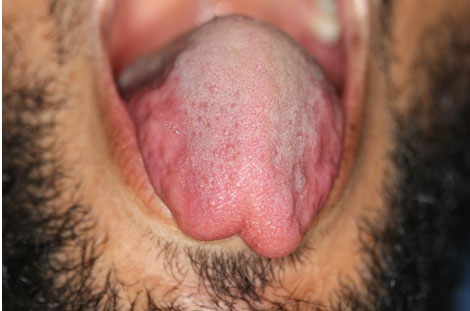
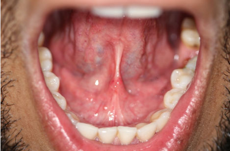
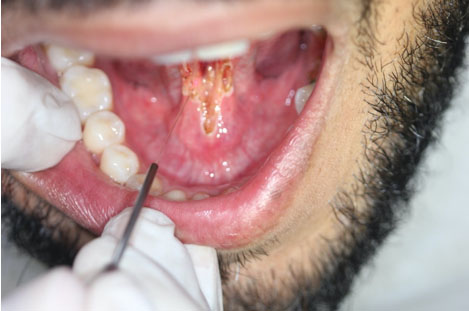
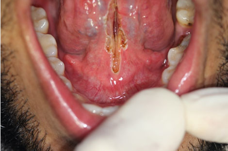
Primarily, the surgeon proceeded intraoral examination confirming the restriction of tongue movements (Figure 1) and limitation in elevation and protrusion of the tongue (Figure 2) in order to confirm ankyloglossia diagnose. Subsequently, infiltrative local anesthesia was administered using 2% Mepivacaine with 1:100,000 adrenaline (Mepiadre, Nova DFL, Brazil) to block pain transmission and ensure adequate local anesthesia. To minimize swelling and optimize surgical conditions, a 3-0 silk suture (Ethicon, Johnson & Johnson, USA) was placed at the tip of the tongue to provide better control during the procedure (Figure 3). This surgical maneuver is essential for better control of the region to be operated and provides security and stability to the surgeon, once this is an anatomically sensitive area near to Wharton’s ducts and the insertion of the frenulum in the floor of the mouth. Then, a scalpel with a #15c blade is used to section the frenulum performing a diamond-shaped pattern in the mucosa (Figure 4). The incision was laterally extended to ensure tongue movements. Using Metzenbaum scissors, the mucosal layer was separated from the underlying muscle layers preventing excessive tension in the suture. The surgeon uses a 6-0 resorbable thread (Vycril, Ethicon, Johnson & Johnson, USA) to place 3 to 7 single knots suture, performing coaptation of the edges of the surgical wound (Figure 5). In no case was it necessary to correct postoperative complications.
Paracetamol (500 mg—every 8 hours in the first 48 hours in case of pain) and chlorhexidine mouthwashes (0.12% chlorhexidine digluconate—3 to 4 times a day for a week) were prescribed. Patients receive orientation for tongue training exercises and dietary guidance for soft and not warm food dietary. The sutures were removed after seven days (Figure 6).
OPERATING PROTOCOL FOR GROUP B
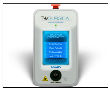
After confirming the limitations of tongue movements due to ankyloglossia (Figure 7 and Figure 8), the infiltrative local anesthesia is proceeded by the surgeon using Mepivacaine hydrochloride 2% with adrenaline 1:100,000 (Mepiadre, Nova DFL, Brazil), in order to isolate local sensibility and Mepivacaine 2% with adrenaline 1:100,000 and block the transmission of pain. With the aid of surgical gauze, the patient’s tongue was gently retracted, positioning the frenulum between the index and middle fingers to avoid deformation and tension. Using a diode laser device (Laser Tw Surgical, MMoptics, Brazil) (Figure 9), the frenulum was sectioned by direct contact of the activated fiber with the target tissue. The incision starts in the insertion area closest to the tip of the tongue and proceeded distally to the oral floor, parallel to the long axis of the tongue. It must be performed on both the right and left sides, with the laser tip in contact mode, using brushing movements. This procedure caused a progressive release of tractions that increased even more with the deepening of the cut towards the midline, allowing an agile lifting of the tongue toward the palate (Figure 10). To allow the procedure to be carried out quickly and accurately, it was necessary to activate the fiber every time the cutting power was inhibited, also combining the removal of residues with gauze moistened in saline solution. The entire procedure must be carried out without irrigation.
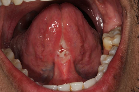
No bleeding was expected, as the use of the surgical laser determines instantaneous coagulation of the mucogingival and connective tissues at the time of the incision. Fiber sectioning was performed to increase tongue mobility. Then, the laser fiber was set in blurred mode, in order to biostimulate the tissues around the treated area and reduce edema and postoperative pain. There is no need to prescribe analgesics or anti-inflammatory drugs. No sutures were necessary, as the laser was able to cauterize the tissue, preventing bleeding immediately after the surgical procedure (Figure 11). Chlorhexidine mouthwashes (0.12% chlorhexidine digluconate—3 to 4 times a day for a week) were prescribed. Patients receive orientation for tongue training exercises and dietary guidance for soft and not warm food dietary. The patients were reassessed 24 h and 7 days after the operation (Figure 12).
POSTOPERATIVE OUTCOMES
All patients demonstrated to perceive an improvement in the movement and elevation of the tongue, and it was possible to observe an objective improvement in the level of tongue mobility in the immediate postoperative period. The patients were reassessed 24 h and 7 days after the operation and no edema or postoperative complications were recorded in any case.


RESULTS
To carry out a comparison between the appraised techniques, two different outcomes were evaluated. For postoperative pain, this paper used the NRS immediately after the surgical procedure and at the review appointment one week after the surgery. In order to assess the quality of wound healing in each group the clinical parameters that make up EHS were measured at a follow-up visit 24 h after the operation. As secondary outcomes, a subjective analysis of trans-surgical bleeding by the operator was performed and average operating time was recorded [12].
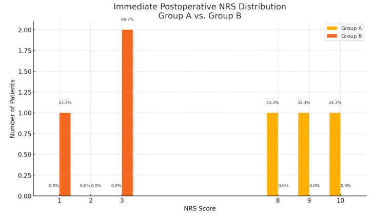
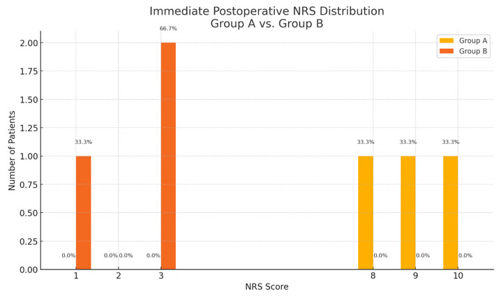
The mean NRS to indicate the level of pain that patients felt immediately after the end of anesthesia in group A patients was 9. Instead, the mean NRS in group B patients at the same time point was 2.32 (Figure 13). Comparison of mean values between groups showed a 74.22% reduction in NRS recorded in the immediate postoperative period in group B compared to group A. Therefore, group A is more prone to more uncomfortable healing, which was confirmed by their NRS recorded in the first postoperative week, with a mean score of 4.33, while patients in group B have a mean NRS score during the first week of 0.33 (Figure 14), so that there was a 92.37% reduction in the NRS recorded in the first postoperative week in group B compared to group A.
The mean EHS for evaluating the quality of wound healing 24 h after the operation in patients in group A was 6.66 (Table 2), while the mean EHS in patients in group B was 10 in all patients evaluated (Table 3), highlighting a mean difference in EHS between the two groups of 33.4%, with the quality of healing in the first 24 h significantly increased in patients in group B. It was observed that in patients in group A, the presence of intraoperative bleeding was recorded, while in patients in group B there was no intraoperative bleeding, improving the view of the surgical site. Finally, the mean operating time for patients in group A was 38.66 min, while the mean operating time for patients in group B was 16.33 min, showing an interesting reduction in intraoperative time.


DISCUSSION
There are specific clinical criteria by which the diagnosis of ankyloglossia can be formulated: during the mouth opening movement the patient will not be able to touch the palate with the tip of the tongue; when performing the protrusion movement the presence of a median sulcus should be noticed; the patient presents a great restriction of the sublingual space; when performing total extrusion, the tongue does not go beyond the vermilion of the lip, causing flexion of the central body of the tongue. Although it is preferable to treat this disease at a very early age, in order to avoid problems with speech ability and orthodontic problems such as lingual inclination of the lower incisors, diastemas and dental rotation, it is also possible to perform these procedures in adult patients who present, along with ankyloglossia, dysphonia problems, nodules on the vocal cords, snoring problems, mandibular disharmony or swallowing-postural syndrome [1],[7],[8],[10],[11],[12],[13]. It is also recommended to ensure that a short frenulum does not cause instability in a mobile prosthesis rehabilitation plan, in which case surgery is strongly recommended, as these procedures can be particularly difficult to perform in patients who have the full form of ankyloglossia.
When compared to the use of diode laser, the conventional method may have some disadvantages during and after surgery. Among the complications related to the surgical technique, we can highlight excessive bleeding, prolonged healing time, and excessive patient discomfort. It is also important to point out that these advantages are also given in terms of surgical results, since laser surgery presents better outcomes when assessing the appearance of keloids, and a lower chance of recurrence [14]. These advantages are because the laser technique has an operating principle, based on tissue interaction, completely different from the classic surgical technique. In fact, the laser not only “cuts” the structures that make up the frenulum (collagen and elastic fiber), but causes great heating (60 °C) and consequently tissue denaturation and coagulation [14]. In fact, the scalpel causes the removal of the edges of the fibrous structures and the elimination of tension, but it keeps both sections intact, provoking an inflammatory reaction in the body that seeks to repair the “damage” [15]. Instead, the denaturation caused by the laser beam causes, first, a significant removal of the fibrous endings caused by both the reduction of physiological traction and the elimination of fibers; then a degradation of the collagen fibers, breaking the weak chemical hydrogen bonds between the collagen chains and facilitating the presence of stronger bonds between the amino acids that organize the structures of proteins (such as lysine). This procedure therefore makes the repair process more difficult for the fibroblasts, which is not immediately viable in the clotted sites, promoting a less favorable environment for the resurgence of the lingual frenulum [15],[16].
The repair process, therefore, is less complex related to laser surgeries, unlike what occurs in the traditional approach, the amount of tissue removed is greater, in addition all coagulated tissue must be removed and only then can new fibers be remodeled, creating a long regeneration process. For this reason, after laser surgeries, the simpler repair process is more evident, with low remodeling of the fibers, great wound contraction, which, together with the non-existent contamination of the surgical site and an extremely fast execution of the procedure itself, allow for less inflammatory exudate, more postoperative comfort, decreasing pain and swelling, and discarding the use of antibiotics [15],[16].
Furthermore, the use of the laser in pulsed mode can guarantee all the specific effects mentioned above and ensure that the tissue is not overheated. In this way, the tissue is prevented from being heated more than necessary, avoiding damage to the surrounding anatomical structures. Finally, the fact is that laser frenectomy can be performed without the use of a scalpel, which decreases bleeding and the need for sutures. Laser frenectomy can be performed in less time with very little discomfort for patients, so that even children are more apt to undergo surgery, facilitating repeat treatment in case of recurrence, due to the absence of a previous traumatic experience [15],[16].
Similar results have been highlighted in other studies [17],[18],[19],[20],[21],[22],[23], making laser-assisted frenectomy a more viable option compared to the conventional surgical method used in recent years. In particular, one study focused on reducing the need for sutures, while other studies highlighted different positive results in diode laser frenectomy when compared to the traditional surgical method, such as reduced patient discomfort, reduced operating time, and reduced need for infiltrative anesthesia [21],[22],[23].
CONCLUSION
Within the limits of this study, the results suggest that laser frenectomy can be considered a better option than the conventional surgical method, especially in patients with problems that prevent them from performing longer-term interventions and patients who are particularly sensitive to pain. The results are encouraging and justify further studies that may include a larger number of subjects, in order to confirm the results obtained on a large scale.
REFERENCES
1.
Nicoloso GF, dos Santos IS, Flores JA, da Silveira BL, Oliveira MDM. An alternative method to treat ankyloglossia. J Clin Pediatr Dent 2016;40(4):319–21. [CrossRef]
[Pubmed]

2.
Fraga MRBA, Barreto KA, Lira TCB, Celerino PRRP, Tavares ITS, de Menezes VA. Anquiloglossia versus amamentação: Qual a evidência de associação? Rev CEFAC 2020;22(3):e12219. [CrossRef]
3.
Procopio, IMS, Costa VPP, Lia EN. Frenotomia lingual em lactentes. Revista Da Faculdade De Odontologia -UPF 2017;22(1).
4.
Varadan M, Chopra A, Sanghavi AD, Sivaraman K, Gupta K. Etiology and clinical recommendations to manage the complications following lingual frenectomy: A critical review. J Stomatol Oral Maxillofac Surg 2019;120(6):549–53. [CrossRef]
[Pubmed]
5.
Lima MPCSL, WR. Anquiloglossia e frenectomia lingual: Relato de caso clínico em adolescente. São Paulo 2009.
6.
Navarro RS, Gontijo I, Haypek P, Eduardo C de P. Lasers em odontopediatria: Estado da arte. Odontopediatria na Prim Infância 2009.
7.
Gontijo I, Navarro RS, Haypek P, Ciamponi AL, Haddad AE. The applications of diode and Er:YAG lasers in labial frenectomy in infant patients. J Dent Child (Chic) 2005;72(1):10–15. [CrossRef]
[Pubmed]
8.
Pompéia LE, Ilinsky RS, Ortolani CLF, Faltin K Júnior. Ankyloglossia and its influence on growth and development of the stomatognathic system. Rev Paul Pediatr 2017;35(2):216–21. [CrossRef]
[Pubmed]
9.
Marchesan IQ, de Castro Martinelli RL, Gusmão RJ. Lingual frenulum: Changes after frenectomy. J Soc Bras Fonoaudiol 2012;24(4):409–12. [CrossRef]
[Pubmed]
10.
Marco CA, Marco AP. Assessment of pain. In: Thomas SH, editor. Emergency department analgesia: An evidence-based guide. Cambridge: Cambridge University Press; 2008. p. 10–18.
11.
Karcioglu O, Topacoglu H, Dikme O, Dikme O. A systematic review of the pain scales in adults: Which to use? Am J Emerg Med 2018;36(4):707–14. [CrossRef]
[Pubmed]
12.
Marini L, Rojas MA, Sahrmann P, Aghazada R, Pilloni A. Early wound healing score: A system to evaluate the early healing of periodontal soft tissue wounds. J Periodontal Implant Sci 2018;48(5):274–83. [CrossRef]
[Pubmed]
13.
Carosi P, Barlattani A Jr, Lorenzi C, Dolci A, Bianchi N, Arcuri C. Diode laser as an adjunct to nonsurgical chronic periodontitis therapy: A review. J Biol Regul Homeost Agents 2020;34(3 Suppl. 1):45–54.
[Pubmed]
14.
Estrin NE, Moraschini V, Zhang Y, Romanos GE, Sculean A, Miron RJ. Combination of Nd:YAG and Er:YAG lasers in non-surgical periodontal therapy: A systematic review of randomized clinical studies. Lasers Med Sci 2022;37(6):2737–43. [CrossRef]
[Pubmed]
15.
Eming SA, Martin P, Tomic-Canic M. Wound repair and regeneration: Mechanisms, signaling, and translation. Sci Transl Med 2014;6(265):265sr6. [CrossRef]
[Pubmed]
16.
Janda P, Sroka R, Mundweil B, Betz CS, Baumgartner R, Leunig A. Comparison of thermal tissue effects induced by contact application of fiber guided laser systems. Lasers Surg Med 2003;33(2):93–101. [CrossRef]
[Pubmed]
17.
Rathnakar B, Rao BSS, Prabhu V, Chandra S, Rai S, Rao ACK, et al. Photo-biomodulatory response of lowpower laser irradiation on burn tissue repair in mice. Lasers Med Sci 2016;31(9):1741–50. [CrossRef]
[Pubmed]
18.
Yadav RK, Verma UP, Sajjanhar I, Tiwari R. Frenectomy with conventional scalpel and Nd:YAG laser technique: A comparative evaluation. J Indian Soc Periodontol 2019;23(1):48–52. [CrossRef]
[Pubmed]
19.
Sarmadi R, Gabre P, Thor A. Evaluation of upper labial frenectomy: A randomized, controlled comparative study of conventional scalpel technique and Er:YAG laser technique. Clin Exp Dent Res 2021;7(4):522–30. [CrossRef]
[Pubmed]
20.
Mazzoni A, Navarro RS, Fernandes KPS, Horliana ACRT, Mesquita-Ferrari RA, Motta PB, et al. Evaluation of the effects of high-level laser and electrocautery in lingual frenectomy surgeries in infants: Protocol for a blinded randomised controlled clinical trial. BMJ Open 2021;11(11):e050733. [CrossRef]
[Pubmed]
21.
Brignardello-Petersen R. Use of lasers seems to result in a small decrease in postoperative complications after labial frenectomy compared with scalpels. J Am Dent Assoc 2020;151(5):e42. [CrossRef]
[Pubmed]
22.
Nammour S. Laser-assisted tongue-tie frenectomy for orthodontic purpose: To suture or not to suture? Photobiomodul Photomed Laser Surg 2019;37(7):381–2. [CrossRef]
[Pubmed]
23.
Viet DH, Ngoc VTN, Anh LQ, Son LH, Chu DT, Ha PTT, et al. Reduced need of infiltration anesthesia accompanied with other positive outcomes in diode laser application for frenectomy in children. J Lasers Med Sci 2019;10(2):92–96. [CrossRef]
[Pubmed]
SUPPORTING INFORMATION
Author Contributions
Carlos Henrique Sardenberg Pereira - Conception of the work, Design of the work, Acquisition of data, Analysis of data, Drafting the work, Revising the work critically for important intellectual content, Final approval of the version to be published, Agree to be accountable for all aspects of the work in ensuring that questions related to the accuracy or integrity of any part of the work are appropriately investigated and resolved.
Otto de Oliveira Magro - Conception of the work, Design of the work, Acquisition of data, Analysis of data, Drafting the work, Revising the work critically for important intellectual content, Final approval of the version to be published, Agree to be accountable for all aspects of the work in ensuring that questions related to the accuracy or integrity of any part of the work are appropriately investigated and resolved.
Vitor Hugo Torres Nunes - Conception of the work, Design of the work, Acquisition of data, Analysis of data, Drafting the work, Revising the work critically for important intellectual content, Final approval of the version to be published, Agree to be accountable for all aspects of the work in ensuring that questions related to the accuracy or integrity of any part of the work are appropriately investigated and resolved.
Marcelo José Pinheir Guedes de Uzeda - Conception of the work, Design of the work, Acquisition of data, Analysis of data, Drafting the work, Revising the work critically for important intellectual content, Final approval of the version to be published, Agree to be accountable for all aspects of the work in ensuring that questions related to the accuracy or integrity of any part of the work are appropriately investigated and resolved.
Sileno Correia Brum - Conception of the work, Design of the work, Acquisition of data, Analysis of data, Drafting the work, Revising the work critically for important intellectual content, Final approval of the version to be published, Agree to be accountable for all aspects of the work in ensuring that questions related to the accuracy or integrity of any part of the work are appropriately investigated and resolved.
Guaranter of SubmissionThe corresponding author is the guarantor of submission.
Source of SupportNone
Consent StatementWritten informed consent was obtained from the patient for publication of this article.
Data AvailabilityAll relevant data are within the paper and its Supporting Information files.
Conflict of InterestAuthors declare no conflict of interest.
Copyright© 2025 Carlos Henrique Sardenberg Pereira et al. This article is distributed under the terms of Creative Commons Attribution License which permits unrestricted use, distribution and reproduction in any medium provided the original author(s) and original publisher are properly credited. Please see the copyright policy on the journal website for more information.