|
Case Report
Conservative management of traumatic pneumocephalus and primary reconstruction following a self-inflicted midfacial gunshot wound: A case report
1 Department of Oral and Maxillofacial Surgery, Moi Teaching and Referral Hospital/Moi University School of Dentistry, Eldoret, Kenya
Address correspondence to:
Dennis Cheruiyot
Department of Oral and Maxillofacial Surgery, Moi Teaching and Referral Hospital, P.O. Box 3, Eldoret 30100,
Kenya
Message to Corresponding Author
Article ID: 100048D01DC2026
Access full text article on other devices

Access PDF of article on other devices


How to cite this article
Cheruiyot D, Rakewa S, Thuku F, Koech L, Wamalwa E. Conservative management of traumatic pneumocephalus and primary reconstruction following a self-inflicted midfacial gunshot wound: A case report. Edorium J Dent 2026;13(2):1–8.ABSTRACT
Introduction: Gunshot wounds to the midface represent a profound surgical challenge integrating acute life-threatening pathophysiology with complex reconstruction. We report the multidisciplinary care of a 52-year-old man who sustained a self-inflicted gunshot injury to the midface, resulting in an extensive panfacial laceration, a dentoalveolar fracture, and minor traumatic pneumocephalus.
Case Report: Immediate care followed Advanced Trauma Life Support (ATLS) principles, prioritizing airway security and hemodynamic resuscitation. Computed tomography (CT) neuroimaging identified frontal lobe pneumocephalus in the absence of tension physiology, prompting a conservative neurosurgical strategy of watchful observation, short-course seizure prophylaxis, and targeted antimicrobial coverage. Operative management comprised thorough wound debridement with high-volume saline irrigation, followed by anatomically layered primary closure using absorbable deep sutures and interrupted non-absorbable monofilament skin sutures. Repeat CT at 24 hours demonstrated near-complete clearance of the intracranial air.
Conclusion: The patient achieved full recovery without neurological sequelae or wound infection, with satisfactory healing evident at the initial postoperative review. This case demonstrates that a structured, protocol-driven approach combining trauma resuscitation, selective non-operative management of intracranial complications, and precise soft tissue reconstruction yields favorable outcomes in high-energy craniofacial ballistic trauma.
Keywords: Ballistic trauma, Facial reconstruction, Gunshot wound, Maxillofacial surgery, Pneumocephalus
INTRODUCTION
Gunshot wounds to the midface represent a profound surgical challenge, integrating acute life-threatening pathophysiology with complex staged reconstruction. Anatomically dense, this region houses critical structures for airway protection, mastication, sensation, and communication. These injuries carry a disproportionately high burden of morbidity relative to their occurrence among firearm-related traumas [1]. Management follows ATLS principles from the outset, with establishment of a secure airway as the foremost priority, followed by hemorrhage control and evaluation for intracranial or cervical spine injury [2].
The ballistic energy transfer in these injuries creates a unique pathophysiology. The anatomical proximity of the paranasal sinuses to the anterior cranial base makes fractures in this region capable of establishing pathways for intracranial injury. Frontal sinus gunshot wounds in particular carry major complication rates above 50%, encompassing cerebrospinal fluid (CSF) fistulae and intracranial sepsis, underscoring the need for watertight dural closure and sinus obliteration during reconstruction [3]. Moreover, the cavitation effect and projectile fragmentation potential create a substantial risk of cerebrovascular injury (CVI). Recent evidence indicates that self-inflicted craniofacial gunshot wounds carry a particularly high incidence of vascular injury. A distinct pattern of blunt CVI-like lesions, often distant from the primary wound tract and initially missed on imaging, has been identified at a rate two to six times higher than in other trauma mechanisms, necessitating a high index of suspicion and systematic radiographic screening to prevent delayed ischemic stroke [4].
Pneumocephalus, the presence of intracranial air, is a cardinal radiological sign of skull base violation. While often managed conservatively when minor and asymptomatic, it necessitates vigilant monitoring for progression to tension pneumocephalus, a neurosurgical emergency characterized by mass effect [5],[6]. The projectile trajectory, particularly an anteriorto-posterior vector, is a critical independent predictor of both vascular injury risk and ultimate neurological outcome [4],[7].
The reconstructive paradigm for these composite defects has matured toward an anatomical subunit approach. Successful restoration of form and function demands a strategic selection from techniques including precise fracture fixation, autogenous bone grafting, local tissue rearrangement, and microvascular free tissue transfer [8]. This reconstruction is inherently staged, beginning with aggressive debridement and initial soft tissue closure while planning for subsequent definitive bony and soft tissue restoration [9].
The management of self-inflicted midfacial gunshot wounds epitomizes the necessity for multidisciplinary coordination between emergency medicine, neurosurgery, and maxillofacial surgery. This case report details the presentation of a 52-year-old male with a severe self-inflicted midfacial gunshot wound resulting in extensive soft tissue injury, dentoalveolar fracture, and minor frontal pneumocephalus. We utilize this case to critically examine key management decisions, focusing on the rationale for conservative neurosurgical management of limited pneumocephalus, the principles guiding thorough debridement and immediate primary layered closure in a ballistic wound, and the evidence-based approach to medical prophylaxis.
CASE REPORT
Pre-Hospital Presentation and Emergency Department Arrival
A 52-year-old male was transported to the Emergency Department via emergency medical services following a reported self-inflicted gunshot wound to the midface. Witnesses reported hearing two separate gunshot reports. On discovery, the patient was conscious and supine in a pool of blood estimated at over 2 L. No clear fluid was observed draining from the patient. Social history identified him as a social consumer of alcohol and a lifelong non-smoker. He was married with four children, all in good health.
Primary and Secondary Survey Findings
On arrival, management was directed by the ATLS framework. The primary survey identified an immediate threat to airway patency due to extensive midfacial soft tissue destruction, active hemorrhage, and aspiration risk. An appropriately sized oropharyngeal airway was inserted to maintain the upper airway, and high-flow oxygen delivered via non-rebreather mask. Two large-bore peripheral intravenous cannulae were placed for fluid resuscitation. Initial observations recorded: blood pressure 118/65 mmHg, core temperature 35.6 °C (mild hypothermia), oxygen saturation 90% on supplemental oxygen, and pulse rate 78 beats per minute. The combination of relative hypotension and estimated blood loss was consistent with compensated hypovolaemic shock.
Once life-threatening conditions were stabilised, the secondary survey proceeded. The patient scored 15 on the Glasgow Coma Scale, with all components intact. Systematic neurological assessment revealed 5/5 motor strength in all limbs and no focal sensory deficits. Cranial nerve testing was limited by the facial injury, though pupillary and gag reflexes appeared preserved. Thoracoabdominal and pelvic examinations were unremarkable. Systemic review found mild conjunctival pallor consistent with acute blood loss, without evidence of jaundice, cyanosis, peripheral oedema, or lymphadenopathy.
Detailed Maxillofacial Examination and Initial Imaging
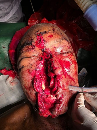
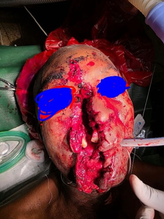
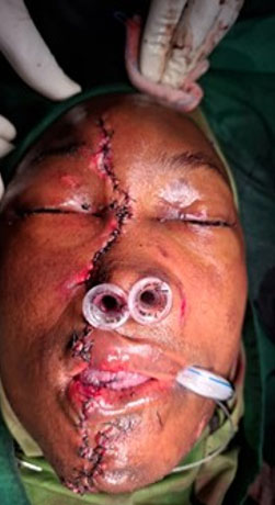
Detailed maxillofacial assessment revealed a severe diagonal laceration traversing the face. Wound morphology indicated the entry point was at the submental or mental midline. The tract extended superiorly through the full thickness of the lower lip, traversed the philtrum and upper lip, deviated laterally to involve the right nasal ala, continued across the right cheek and infraorbital region, and terminated at the right frontal bone near the anterior hairline (Figure 1A). Wound edges were ragged and bruised, with visible embedded debris and gross contamination. Palpation confirmed bony exposure at the anterior maxilla and inferior orbital rim. Examination revealed significant mucosal tearing, a disrupted gingivobuccal sulcus, and mobility of the anterior maxillary alveolar segment, clinically diagnostic of a dentoalveolar fracture involving at least teeth 12 and 21 (maxillary left lateral incisor and right central incisor) (Figure 1).
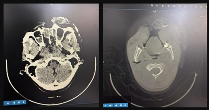
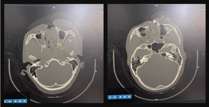
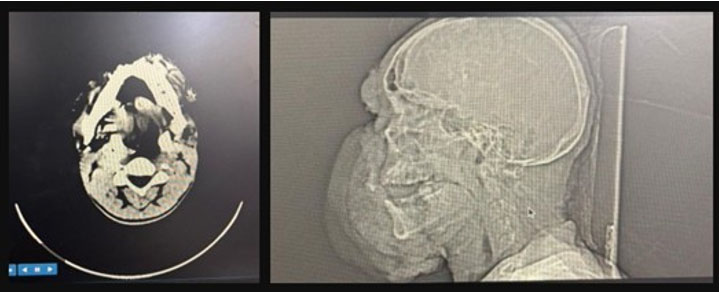
Given the high-velocity mechanism and trajectory suggesting possible intracranial extension, an urgent non-contrast computed tomography (CT) scan of the head and maxillofacial skeleton was obtained. Computed tomography findings were pivotal for subsequent management. First, it confirmed the presence of a small volume of pneumocephalus, localized to the right frontal lobe parenchyma (Figure 1B and Figure 1C). This radiographic finding is definitive evidence of communication between the facial injury and the intracranial space, likely via a fracture through the frontal sinus floor or the orbital roof. Second, the CT scan precisely delineated the dentoalveolar fracture as extending from the region of tooth 12 to tooth 22, involving the entire anterior maxillary alveolar process. No other skull fractures or intracranial hemorrhages were identified (Figure 2, Figure 3, Figure 4).
Multidisciplinary Decision Making and Preoperative Medical Management
An urgent multidisciplinary case conference was convened involving emergency medicine, neurosurgery, and maxillofacial surgery.
Concurrently, the maxillofacial surgical team formulated a medical and operative plan. A short, targeted antibiotic course was commenced to address the anticipated polymicrobial inoculum from the oropharyngeal and sinus flora, comprising intravenous ceftriaxone (1 g twice daily) and intravenous metronidazole (400 mg three times daily) for five days. This regimen is consistent with current evidence-based guidance on perioperative antimicrobial prophylaxis in open facial fractures. Pain control was achieved using scheduled intravenous paracetamol (1 g three times daily), with intravenous tramadol (100 mg twice daily) available for breakthrough analgesia.
Operative Procedure: Surgical Debridement and Reconstruction
The patient was transferred to the operating theatre for urgent wound management. Full adherence to the World Health Organization surgical safety checklist was confirmed. General anesthesia was induced with intravenous propofol, and neuromuscular blockade was achieved with atracurium to facilitate airway security via endotracheal intubation. Anesthesia was maintained with 1.2% isoflurane in an oxygen-air mixture. Intravenous dexamethasone (8 mg) was given to attenuate perioperative inflammatory edema.
Preoperative Preparation and Initial Wound Assessment
The patient was placed supine. Skin preparation was performed meticulously. An initial rinse with sterile water and mild soap cleared gross surface contamination, with the eyes protected using sterile ophthalmic ointment and adhesive drapes. This was followed by a standardized surgical scrub using a chlorhexidine gluconate-based solution, applied widely from the scalp to the clavicles. The patient was then draped in a sterile fashion, exposing the entire facial injury (Figure 5).
Surgical Debridement
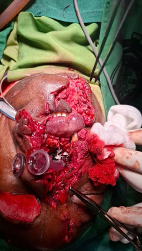
Radical debridement principles guided this operative phase. All clearly non-viable tissue was sharply excised using fine forceps and a scalpel, including contused skin margins, devitalised subcutaneous fat, and fragmented muscle fibers. Thorough removal of all embedded foreign material, including bullet fragments, soil particles, and other debris, was prioritized. Following excision, the wound was subjected to high-volume pulsatile irrigation. Normal saline (0.9% NaCl) was delivered under pressure via a large syringe and irrigation catheter, systematically reaching all recesses of the wound tract. The total irrigation volume exceeded 5 L, consistent with recommended practice for contaminated ballistic wounds to achieve mechanical bacterial load reduction. Hemostasis was secured with targeted bipolar electrocautery to limit thermal injury to adjacent viable structures.
Layered Anatomical Closure
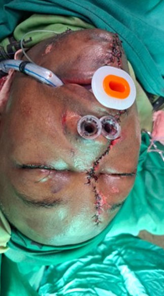
Following complete debridement and irrigation, the wound bed consisted of clean, bleeding tissue margins with preserved vascularity, permitting a decision for immediate primary closure. The reconstruction was performed by two surgeons to ensure precision and efficiency. The closure was executed in a layered fashion. First, the orbicularis oris muscle fibers in the lip were meticulously reapproximated using interrupted 4-0 polyglactin 910 (Vicryl) sutures to restore the oral sphincter’s functional continuity. Similarly, the subcutaneous tissue and fascial layers in the cheek and forehead were closed with interrupted 3-0 Vicryl sutures. This deep layer closure is fundamental for eliminating dead space, reducing tension on the skin closure, and providing a scaffold for healing. Subsequently, the dermal layer was closed with interrupted 5-0 Vicryl sutures. Finally, the skin was closed using simple interrupted 5-0 nylon sutures (Figures 6, Figure 7, Figure 8). The use of a nonabsorbable monofilament suture for the skin minimizes tissue reaction and the risk of suture sinus formation, while the interrupted technique permits selective removal of sutures if a localized infection develops.
Postoperative Management and Clinical Follow-Up
The patient was extubated in theatre following complete reversal of neuromuscular blockade and transferred to the Surgical Intensive Care Unit (SICU) for close neurological and respiratory surveillance. Thirty-degree head-of-bed elevation was maintained to promote venous drainage and facilitate resolution of the pneumocephalus. Neurological observations were conducted hourly in the early postoperative period. Repeat CT head at 24 hours confirmed near-complete resolution of the right frontal pneumocephalus with no new intracranial findings, validating the conservative neurosurgical strategy.
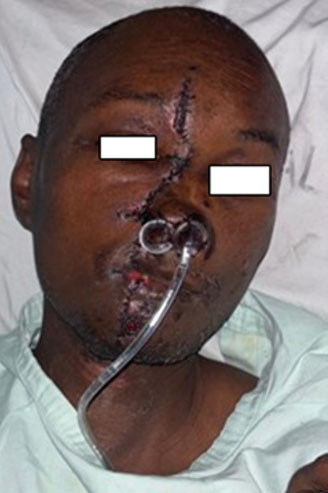
The maxillofacial wounds were inspected daily. By the fifth postoperative day, the surgical sites exhibited excellent healing (Figure 9). The nylon sutures were intact, the surrounding skin showed only mild erythema, and there was no sign of infection, hematoma, or wound dehiscence. The significant facial edema presents postoperatively had markedly subsided. The patient was transitioned from intravenous to oral antibiotics (amoxicillin-clavulanate 625 mg twice daily for five days) and oral analgesics. Oral intake was advanced from clear liquids to a soft diet as tolerated.
Psychosocial and Ethical Considerations
Given the nature of the injury, a formal psychiatric consultation was obtained prior to discharge. The patient was diagnosed with a major depressive episode and commenced on appropriate pharmacotherapy with a plan for outpatient psychological follow-up. Furthermore, in strict adherence to ethical standards for publication, a detailed conversation was held with the patient regarding the use of his de-identified clinical data and photographs for educational purposes. Written informed consent was obtained, documented in the medical record, and filed separately, complying with international guidelines for case report publication. The patient was discharged home on postoperative day seven in a stable condition with arranged follow-up in both the maxillofacial surgery and psychiatry clinics.
DISCUSSION
This case of a self-inflicted craniofacial gunshot injury serves as a useful illustration of the structured approach required for high-energy facial trauma. The favorable clinical result highlights the value of combining prompt resuscitation with systematic complication surveillance and technically sound surgical repair. The wounding trajectory, which involved the anterior maxilla, nasal skeleton, and frontal region, categorized this patient as high-risk for both acute and delayed complications, necessitating judicious clinical decision-making at each stage of management.
The immediate priority of securing a tenuous airway was appropriately addressed, aligning with the foundational principle of ATLS. The subsequent identification of frontal lobe pneumocephalus on CT represented a critical juncture in management. The decision to pursue conservative neurosurgical management was predicated on the patient’s intact neurological examination and the absence of tension physiology on imaging. This approach is strongly supported by literature which advocates for nonoperative observation in cases of asymptomatic, minor pneumocephalus, reserving surgical intervention for scenarios involving mass effect or neurological decline [10],[11]. However, this case also highlights a significant risk associated with the anterior-to-posterior projectile trajectory. Recent evidence specific to craniofacial self-inflicted gunshot wounds indicates a substantially elevated incidence of associated cerebrovascular injury, including vessel dissection, which can be clinically silent initially [4]. While our patient remained neurologically stable, this literature underscores the necessity of a high index of suspicion and consideration for advanced vascular imaging in similar injury patterns to prevent catastrophic delayed stroke.
The surgical strategy of aggressive debridement followed by immediate layered primary closure was executed successfully. The utilization of copious irrigation exceeding 5 L of normal saline is a recommended practice to reduce microbial load in contaminated ballistic wounds. The choice of definitive primary closure was contingent upon achieving a wound bed of viable tissue following debridement. As reported by Soto et al., gunshot injuries to the frontal sinus are associated with major complication rates exceeding 50%, frequently requiring advanced reconstructive techniques such as cranialization supported by local or free tissue flaps to achieve a durable repair [3]. In the present case, although the frontal bone was involved, there was no frontal sinus violation of sufficient magnitude to necessitate such complex reconstruction, enabling a more straightforward operative approach.
The layered closure technique, employing absorbable sutures for deep structural layers and a non-absorbable monofilament for the skin, effectively served both functional and aesthetic objectives. By eliminating dead space, re-establishing muscular continuity, and enabling precise, low-tension skin apposition, this method is consistent with the anatomic subunit philosophy of composite facial reconstruction, which prioritizes individual restoration of each structural and soft tissue component [8]. Where greater composite defects with osseous loss are encountered, the reconstructive algorithm escalates to encompass bone grafting and microvascular free tissue transfer, modalities that would inform any subsequent staged rehabilitation for this patient [8],[9].
Current guidance increasingly favors levetiracetam over phenytoin where the former is available [12],[13].
Comprehensive care in such cases necessarily extends beyond the surgical field. The need for timely psychiatric assessment in deliberate self-harm presentations is well established, and informed consent for publication was obtained in accordance with the CARE guidelines for case report ethics [14],[15]. In conclusion, this case affirms that optimal outcomes in complex midfacial ballistic trauma require a coordinated, multidisciplinary approach underpinned by rigorous adherence to trauma protocols, vigilant recognition of potential complications, technically precise surgical management, and comprehensive patient-centered care.
CONCLUSION
This report documents the successful management of a complex self-inflicted craniofacial gunshot injury using a structured, multidisciplinary protocol. The favorable outcome was realized through strict adherence to the principles of airway protection and hemorrhage control, evidence-based medical management of the intracranial injury, and meticulous surgical debridement with layered primary wound closure. Conservative neurosurgical management of the minor pneumocephalus was validated by near-complete radiological clearance at 24 hours, while the reconstructive approach conformed to contemporary standards for ballistic facial trauma. This case reinforces that optimal outcomes in high-energy facial injury depend on systematic resuscitation, vigilant surveillance for occult vascular injury, precise anatomical repair, and holistic care encompassing both physical and psychological rehabilitation.
REFERENCES
1.
Kaufman Y, Cole P, Hollier LH Jr. Facial gunshot wounds: Trends in management. Craniomaxillofac Trauma Reconstr 2009;2(2):85–90. [CrossRef]
[Pubmed]

2.
Norris O, Mehra P, Salama A. Maxillofacial gunshot injuries at an urban level I trauma center—10-year analysis. J Oral Maxillofac Surg 2015;73(8):1532–9. [CrossRef]
[Pubmed]
3.
Soto E, Ovaitt AK, Clark AR, Tindal RR, Chiasson KF, Aryanpour Z, et al. Reconstructive management of gunshot wounds to the frontal sinus: An urban trauma center’s perspective. Ann Plast Surg 2021;86(6S Suppl 5):S550–4. [CrossRef]
[Pubmed]
4.
Yoon J, Gebran S, Elegbede A, Day S, Wasicek P, Liang Y, et al. Blunt cerebrovascular injury-like injury observed in patients with craniofacial self-inflicted gunshot wounds. J Craniofac Surg 2022;33(4):1046–50. [CrossRef]
[Pubmed]
5.
Jundt JS, Bohnen A, Galbraith BS, Melville J. Tension pneumocephalus: Case report and review. Int J Oral Maxillofac Surg 2022;51(12):1570–2. [CrossRef]
[Pubmed]
6.
Śmiłowska K, Sznajder-Stacha K, Kocyłowski D, Popek A, Rozpondek K, Grechuta M, et al. Pneumocephalus as a rare complication: A systematic review plus clinical vignette. Neurol Neurochir Pol 2021;55(3):253–68. [CrossRef]
[Pubmed]
7.
Lazaridis C, Mansour A, Alvarado-Dyer R, Tangonan R, Loggini A, Kramer C, et al. Civilian firearm-inflicted brain injury: Coagulopathy, vascular injuries, and triage. Curr Neurol Neurosci Rep 2021;21(9):47. [CrossRef]
[Pubmed]
8.
Heffern E, Nevil C, Przylecki W, Andrews BT. Anatomic subunit approach to composite reconstruction of facial gunshot wounds. J Craniofac Surg 2021;32(7):2487–90. [CrossRef]
[Pubmed]
9.
Vaca EE, Bellamy JL, Sinno S, Rodriguez ED. Management of high-energy avulsive ballistic facial injury: A review of the literature and algorithmic approach. Plast Reconstr Surg Glob Open 2018;6(3):e1693. [CrossRef]
[Pubmed]
10.
Krishnan K, Paterson S, Adhiyaman V. Pneumocephalus. QJM 2011;104(12):1097. [CrossRef]
[Pubmed]
11.
Dabdoub CB, Salas G, Silveira Edo N, Dabdoub CF. Review of the management of pneumocephalus. Surg Neurol Int 2015;6:155. [CrossRef]
[Pubmed]
12.
Frontera JA, Gilmore EJ, Johnson EL, Olson D, Rayi A, Tesoro E, et al. Guidelines for seizure prophylaxis in adults hospitalized with moderate-severe traumatic brain injury: A clinical practice guideline for health care professionals from the Neurocritical Care Society. Neurocrit Care 2024;40(3):819–44. [CrossRef]
[Pubmed]
13.
Riley DS, Barber MS, Kienle GS, Aronson JK, von Schoen-Angerer T, Tugwell P, et al. CARE guidelines for case reports: Explanation and elaboration document. J Clin Epidemiol 2017;89:218–35. [CrossRef]
[Pubmed]
14.
Angriman F, Taran S, Angeloni N, Devion C, Lee JW, Adhikari NKJ. Antiseizure medications in adult patients with traumatic brain injury: A systematic review and Bayesian network meta-analysis. Crit Care Explor 2024;6(10):e1160. [CrossRef]
[Pubmed]
15.
Coccolini F, Sartelli M, Sawyer R, Rasa K, Ceresoli M, Viaggi B, et al. Antibiotic prophylaxis in injury: Global Alliance for Infection in Surgery, Surgical Infection Society Europe, World Surgical Infection Society, American Association for the Surgery of Trauma, and World Society of Emergency Surgery guidelines. J Trauma Acute Care Surg 2024;96(4):674–82. [CrossRef]
[Pubmed]
SUPPORTING INFORMATION
Acknowledgments
The authors wish to acknowledge the nursing and ancillary staff of Moi Teaching and Referral Hospital whose dedicated care contributed to this patient’s recovery.
Artificial intelligence (AI) use in the article: No generative AI technology was used in the preparation, writing, or analysis of this manuscript.
Dennis Cheruiyot - Conception of the work, Design of the work, Acquisition of data, Analysis of data, Drafting the work, Revising the work critically for important intellectual content, Final approval of the version to be published, Agree to be accountable for all aspects of the work in ensuring that questions related to the accuracy or integrity of any part of the work are appropriately investigated and resolved.
Shem Rakewa - Conception of the work, Design of the work, Acquisition of data, Analysis of data, Drafting the work, Revising the work critically for important intellectual content, Final approval of the version to be published, Agree to be accountable for all aspects of the work in ensuring that questions related to the accuracy or integrity of any part of the work are appropriately investigated and resolved.
Francis Thuku - Conception of the work, Design of the work, Acquisition of data, Analysis of data, Drafting the work, Revising the work critically for important intellectual content, Final approval of the version to be published, Agree to be accountable for all aspects of the work in ensuring that questions related to the accuracy or integrity of any part of the work are appropriately investigated and resolved.
Lionel Koech - Conception of the work, Design of the work, Acquisition of data, Analysis of data, Drafting the work, Revising the work critically for important intellectual content, Final approval of the version to be published, Agree to be accountable for all aspects of the work in ensuring that questions related to the accuracy or integrity of any part of the work are appropriately investigated and resolved.
Exavier Wamalwa - Conception of the work, Design of the work, Acquisition of data, Analysis of data, Drafting the work, Revising the work critically for important intellectual content, Final approval of the version to be published, Agree to be accountable for all aspects of the work in ensuring that questions related to the accuracy or integrity of any part of the work are appropriately investigated and resolved.
Guaranter of SubmissionThe corresponding author is the guarantor of submission.
Source of SupportNone
Consent StatementWritten informed consent was obtained from the patient for publication of this article.
Data AvailabilityAll relevant data are within the paper and its Supporting Information files.
Conflict of InterestAuthors declare no conflict of interest.
Copyright© 2026 Dennis Cheruiyot et al. This article is distributed under the terms of Creative Commons Attribution License which permits unrestricted use, distribution and reproduction in any medium provided the original author(s) and original publisher are properly credited. Please see the copyright policy on the journal website for more information.